
Hypoxemic Respiratory Failure human body is deprived of its most essential element, the consequences cascade through every system with devastating precision. Oxygen, the invisible fuel that powers our cellular engines, becomes a precious commodity when the lungs fail to perform their primary function. Hypoxemic respiratory failure represents one of the most critical conditions encountered in modern medicine. In this state, the blood simply cannot maintain adequate oxygen levels despite the body’s desperate attempts to compensate. This condition does not discriminate, affecting patients across all age groups and from every walk of life, transforming routine breathing into a monumental struggle for survival.
The journey into understanding hypoxemic respiratory failure begins with appreciating the delicate balance that maintains life itself. Every breath we take initiates a complex series of events that ultimately deliver oxygen to mitochondria, the powerhouses of our cells. When this pathway is disrupted, whether by disease, injury, or environmental factors, the body’s response is swift and often insufficient. The medical community has made tremendous strides in recognizing and treating this condition, yet it remains a leading cause of morbidity and mortality in intensive care units worldwide. This comprehensive exploration will delve into the mechanisms, manifestations, and management strategies that define our current understanding of this life-threatening condition.
The Fundamental Nature of Hypoxemic Respiratory Failure
At its core, hypoxemic respiratory failure represents a failure of gas exchange within the lungs, specifically the inability to adequately oxygenate arterial blood. This condition is formally defined as a partial pressure of arterial oxygen below sixty millimeters of mercury while the patient is breathing room air at sea level. What makes this condition particularly dangerous is its potential to develop silently, with patients often unaware of their deteriorating oxygen status until significant damage has occurred. The lungs, which normally serve as the body’s primary oxygen exchange interface, become inefficient or ineffective at transferring oxygen from the alveoli into the pulmonary capillary blood.
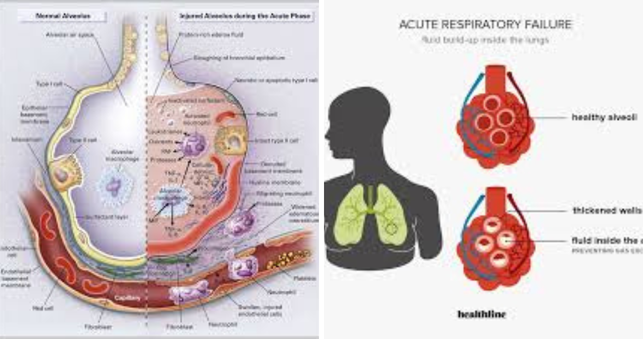
The pathophysiology underlying this condition involves multiple potential mechanisms that can act independently or in concert. The most common causes include ventilation-perfusion mismatch, where areas of the lung receive blood flow without adequate ventilation or vice versa. Intrapulmonary shunting occurs when blood passes through the lungs without participating in gas exchange, essentially bypassing the oxygenation process entirely. Diffusion impairment, though less common, can also contribute when the alveolar-capillary membrane becomes thickened or damaged, preventing efficient oxygen transfer. Understanding these fundamental mechanisms provides the foundation for recognizing why hypoxemic respiratory failure demands such urgent medical attention.
Recognizing the Warning Signs and Clinical Presentation
The clinical presentation of hypoxemic respiratory failure can vary dramatically depending on the underlying cause and the rapidity of onset. Patients typically present with the classic triad of dyspnea, tachypnea, and increasing anxiety as their bodies struggle to maintain adequate oxygen delivery. The visible signs of respiratory distress often include the use of accessory muscles, nasal flaring, and retractions in more severe cases. Cyanosis, perhaps the most recognizable sign of significant hypoxemia, manifests as a bluish discoloration of the skin and mucous membranes when deoxygenated hemoglobin reaches critical levels in the peripheral circulation.
Beyond these visible manifestations, patients frequently experience a constellation of symptoms that can be subtle initially but become increasingly pronounced as the condition worsens. Confusion and altered mental status often develop as cerebral oxygenation declines, with patients becoming agitated or somnolent depending on the severity of their oxygen debt. Tachycardia and hypertension frequently accompany the body’s sympathetic response to hypoxemia, while diaphoresis and pallor reflect the systemic stress of the condition. It is crucial to recognize that the absence of cyanosis does not rule out significant hypoxemia, particularly in patients with anemia where hemoglobin levels are insufficient to produce visible color changes.
The Complex Pathophysiology Behind Oxygen Deprivation
Understanding the mechanisms that drive hypoxemic respiratory failure requires a detailed examination of pulmonary physiology and the factors that can disrupt normal gas exchange. Ventilation-perfusion mismatch represents the most common underlying problem, occurring when some regions of the lung receive ventilation but not perfusion, or perfusion without adequate ventilation. This mismatch can result from conditions such as pneumonia, where consolidated lung regions remain perfused but not ventilated, or from pulmonary embolism, where ventilated regions lose their blood supply. The body attempts to compensate through hypoxic vasoconstriction, redirecting blood flow to better-ventilated areas, but this mechanism has its limits.
Shunting represents a more severe form of gas exchange failure where blood physically bypasses ventilated areas of the lung, effectively preventing any gas exchange from occurring. This can occur anatomically through congenital defects or functionally through conditions such as atelectasis or severe pneumonia where entire regions of the lung become nonfunctional. The shunt fraction, representing the percentage of cardiac output that fails to participate in gas exchange, directly correlates with the severity of hypoxemia. Unlike other causes of hypoxemia, shunting responds poorly to supplemental oxygen therapy because the blood that bypasses the lungs never has the opportunity to become oxygenated regardless of inspired oxygen concentration.
Hypoxemic Respiratory Failure Causes and Risk Factors
The etiologies of hypoxemic respiratory failure span a broad spectrum of conditions affecting both the respiratory and cardiovascular systems. Acute respiratory distress syndrome stands as one of the most severe causes, characterized by diffuse alveolar damage leading to increased permeability of the alveolar-capillary membrane. This condition, often triggered by sepsis, pneumonia, or trauma, results in protein-rich fluid accumulating in the alveoli, severely impairing gas exchange. The resulting hypoxemia is frequently refractory to conventional oxygen therapy, requiring mechanical ventilation and often prone positioning to improve oxygenation.
Pneumonia, whether community-acquired or hospital-acquired, represents another major cause of hypoxemic respiratory failure. The inflammatory response to infection leads to alveolar consolidation and exudate formation, creating areas of ventilation-perfusion mismatch and shunting. Patients with underlying chronic lung disease, particularly chronic obstructive pulmonary disease and interstitial lung disease, face increased risk due to their reduced respiratory reserve and impaired baseline gas exchange. Pulmonary edema, whether cardiogenic or non-cardiogenic, creates another pathway to hypoxemia through fluid accumulation in the alveolar spaces, increasing diffusion distance and creating regions of mismatch.
Diagnostic Approaches and Clinical Assessment
The diagnosis of hypoxemic respiratory failure begins with a thorough clinical assessment incorporating patient history, physical examination, and appropriate diagnostic testing. Arterial blood gas analysis remains the gold standard for confirming the diagnosis, providing objective measurements of arterial oxygen tension, carbon dioxide levels, and acid-base status. The calculated alveolar-arterial gradient offers valuable information about the efficiency of gas exchange, helping to differentiate between hypoxemia caused by respiratory failure versus other causes such as decreased inspired oxygen concentration or hypoventilation.
Pulse oximetry serves as a vital noninvasive monitoring tool, providing continuous assessment of oxygen saturation that helps guide therapeutic decisions. However, clinicians must remain aware of the limitations of pulse oximetry, including potential inaccuracies in patients with poor perfusion, carbon monoxide poisoning, or certain hemoglobinopathies. Chest imaging, particularly computed tomography, provides essential information about the distribution and nature of pulmonary pathology, helping to identify treatable causes such as pneumothorax, pleural effusion, or infiltrative disease. Echocardiography may be necessary to assess cardiac function and exclude cardiogenic causes of pulmonary edema, which can mimic primary respiratory failure.
The Role of Ventilation-Perfusion Relationships
The concept of ventilation-perfusion matching forms the cornerstone of understanding normal gas exchange and its disruption in hypoxemic respiratory failure. In a healthy lung, ventilation and perfusion are carefully matched to optimize oxygen uptake and carbon dioxide elimination. The normal ventilation-perfusion ratio varies throughout the lung, with gravitational effects causing relative differences between dependent and nondependent regions. When this delicate balance is disturbed, whether through regional airway obstruction, vascular changes, or parenchymal disease, the efficiency of gas exchange deteriorates.
The body employs several compensatory mechanisms to address ventilation-perfusion mismatch, most notably hypoxic pulmonary vasoconstriction, which diverts blood flow away from poorly ventilated regions toward better-ventilated areas. This response, mediated by local factors including nitric oxide and endothelin, represents a remarkable adaptive mechanism that maintains oxygenation despite regional abnormalities. However, these compensatory mechanisms can be overwhelmed, particularly in widespread disease processes, leading to clinically significant hypoxemia. Understanding the dynamics of ventilation-perfusion relationships informs therapeutic decisions, including the appropriate use of supplemental oxygen, positive end-expiratory pressure, and positional therapy.
Treatment Strategies and Oxygen Therapy
The management of hypoxemic respiratory failure begins with the administration of supplemental oxygen, delivered through various devices depending on the patient’s oxygen requirements and clinical stability. Nasal cannula provides low-flow oxygen delivery suitable for patients with mild to moderate hypoxemia, while high-flow nasal cannula systems can deliver heated, humidified oxygen at flow rates sufficient to meet the demands of more severely compromised patients. Noninvasive ventilation, including continuous positive airway pressure and bilevel positive airway pressure, offers significant advantages in appropriate patients by providing positive pressure support that improves oxygenation and reduces the work of breathing.
When noninvasive strategies prove insufficient, invasive mechanical ventilation becomes necessary to support gas exchange and protect the airway. The selection of appropriate ventilator settings requires careful consideration of the underlying pathophysiology, with strategies such as lung-protective ventilation incorporating low tidal volumes and appropriate positive end-expiratory pressure to minimize ventilator-associated lung injury. Prone positioning has emerged as a valuable adjunctive therapy for patients with severe hypoxemic respiratory failure, particularly those with ARDS, by improving ventilation-perfusion matching and reducing the effects of gravitational forces on lung mechanics. Extracorporeal membrane oxygenation represents the most advanced form of respiratory support, serving as a salvage therapy for patients with refractory hypoxemia despite optimal conventional management.
Complications and Systemic Effects
The systemic consequences of hypoxemic respiratory failure extend far beyond the respiratory system, affecting nearly every organ system in the body. The cardiovascular system bears a significant burden, with the heart required to work harder to maintain tissue oxygen delivery while often struggling with reduced venous return and increased pulmonary vascular resistance. Myocardial ischemia, arrhythmias, and right ventricular dysfunction can develop as the condition progresses, particularly in patients with underlying cardiac disease. The resulting hemodynamic instability frequently complicates management, creating a complex interplay between respiratory and cardiovascular dysfunction.
Neurologic manifestations range from subtle cognitive changes to profound alterations in consciousness, reflecting the brain’s extreme sensitivity to oxygen deprivation. Patients may develop confusion, agitation, or conversely, somnolence and lethargy as cerebral oxygenation deteriorates. In severe cases, seizures or focal neurologic deficits may occur, mimicking primary neurologic conditions. The gastrointestinal system may also suffer, with splanchnic vasoconstriction leading to mucosal ischemia and the potential for bacterial translocation. Renal function may decline, particularly in the context of systemic hypotension or the use of nephrotoxic medications necessary for management. The development of multiple organ dysfunction significantly worsens prognosis, emphasizing the importance of early recognition and aggressive treatment.
The Role of Positional Therapy
Positional therapy has emerged as a crucial component in managing hypoxemic respiratory failure, particularly in patients with acute respiratory distress syndrome. Prone positioning, in which patients are placed face-down for extended periods, has demonstrated significant benefit in improving oxygenation and reducing mortality in carefully selected patients. The mechanisms underlying this benefit include improved ventilation-perfusion matching, more homogeneous distribution of alveolar ventilation, and reduced compression of dependent lung regions. This simple intervention, when properly implemented, can dramatically improve oxygenation and reduce the need for high levels of ventilatory support.
Beyond prone positioning, other positional strategies can be beneficial in specific clinical scenarios. The sitting or semi-recumbent position improves diaphragmatic excursion and reduces the work of breathing in many patients, while also decreasing the risk of ventilator-associated pneumonia. Lateral positioning may be beneficial in patients with asymmetric disease processes, allowing gravity to improve ventilation to affected areas. In patients with unilateral lung disease, positioning the healthy lung dependent can improve oxygenation by promoting better blood flow to ventilated regions. These positional strategies, while seemingly straightforward, require careful implementation and monitoring to maximize benefit while minimizing potential complications.
The Importance of Nutritional Support
The metabolic demands imposed by hypoxemic respiratory failure are substantial, making nutritional support an essential component of comprehensive management. The increased work of breathing, combined with the systemic inflammatory response, can lead to rapid depletion of energy stores and loss of lean body mass. Early initiation of enteral nutrition, when feasible, is preferred to maintain gut integrity and reduce the risk of bacterial translocation. For patients requiring mechanical ventilation, careful attention to caloric and protein requirements helps preserve respiratory muscle function and facilitate eventual weaning from ventilatory support.
Specific nutritional considerations in the setting of hypoxemic respiratory failure include the management of carbohydrate and fat metabolism. High carbohydrate loads can increase carbon dioxide production, potentially worsening respiratory acidosis, while appropriate fat supplementation can provide calories without significantly increasing the respiratory quotient. The delivery of micronutrients, particularly phosphorus, magnesium, and zinc, is critical for maintaining adequate respiratory muscle function. Electrolyte imbalances, which frequently occur in the setting of critical illness, must be carefully monitored and corrected to prevent cardiac arrhythmias and optimize respiratory muscle contractility. The nutritional plan should be individualized based on the patient’s baseline nutritional status, underlying comorbidities, and degree of metabolic stress.
The Psychological Impact and Patient Experience
The experience of hypoxemic respiratory failure extends far beyond the physiological manifestations, profoundly affecting patients’ psychological well-being and quality of life. The sensation of air hunger, perhaps the most distressing symptom, can trigger severe anxiety and panic, potentially exacerbating the underlying respiratory dysfunction. Patients frequently describe the experience as terrifying, feeling as though they are suffocating despite receiving what appears to be adequate medical support. The psychological trauma associated with respiratory failure can persist long after the acute episode has resolved, contributing to the development of post-traumatic stress disorder and other mental health conditions.
The impact on families and caregivers should not be underestimated, as witnessing a loved one struggle to breathe can be profoundly distressing. The uncertainty surrounding prognosis, the complexity of medical interventions, and the potential for long-term disability create significant emotional burden. Effective communication between healthcare providers, patients, and families is essential in managing these psychological aspects of care. The integration of palliative care principles, including symptom management and goals of care discussions, can significantly improve the patient and family experience while ensuring that treatment aligns with patient values and preferences. The recognition of psychological needs as integral components of comprehensive care represents an important advance in managing this complex condition.
Long-Term Outcomes and Recovery
The trajectory of recovery from hypoxemic respiratory failure varies dramatically based on the underlying cause, severity of illness, and patient-specific factors. While some patients recover completely, regaining normal respiratory function and quality of life, others experience persistent impairments that may require ongoing medical support and rehabilitation. Patients who required mechanical ventilation often face prolonged recovery periods, with many describing significant limitations in physical function and exercise tolerance for months or years after the acute episode. The development of post-intensive care syndrome, encompassing physical, cognitive, and psychological impairments, highlights the long-term consequences that can result from critical illness.
The rehabilitation process begins during the acute phase of illness, with early mobilization and physical therapy playing essential roles in preserving muscle function and preventing complications. As patients recover, progressive exercise training and pulmonary rehabilitation can improve functional capacity and quality of life. For patients with persistent respiratory impairment, home oxygen therapy may be necessary to maintain adequate oxygenation during activities of daily living. The prevention of recurrent episodes is paramount, with careful attention to smoking cessation, vaccination, and management of underlying chronic conditions. The longitudinal care of patients who have survived hypoxemic respiratory failure requires coordination among multiple healthcare providers and a patient-centered approach that addresses the full spectrum of recovery needs.
The Role of Precision Medicine
The emerging field of precision medicine offers promising opportunities to improve outcomes in patients with hypoxemic respiratory failure by tailoring treatment to individual patient characteristics. Genetic factors may influence susceptibility to various causes of respiratory failure as well as response to specific interventions. Inflammatory biomarkers can help identify patients at highest risk for poor outcomes or those most likely to benefit from specific therapies. The integration of sophisticated monitoring techniques, including continuous assessment of respiratory mechanics and hemodynamics, enables real-time adjustment of therapeutic interventions to optimize patient outcomes.
Molecular and genomic profiling may eventually guide the selection of targeted therapies for conditions such as acute respiratory distress syndrome, where substantial heterogeneity in pathophysiology complicates clinical management. The development of artificial intelligence and machine learning algorithms holds promise for identifying patterns of disease progression and predicting response to specific treatments. These advances, while still in early stages of application, represent the future of respiratory medicine, moving away from protocol-based management toward individualized care that accounts for the unique characteristics of each patient’s condition. The challenge lies in translating these technological advances into practical clinical tools that can improve outcomes in diverse healthcare settings.
The Global Burden of Respiratory Failure
The impact of hypoxemic respiratory failure extends far beyond individual patients, representing a significant global health challenge with substantial economic and social consequences. In developed countries, the incidence of respiratory failure requiring intensive care continues to rise, driven by an aging population and increasing prevalence of chronic diseases. The high cost of intensive care, combined with the potential for long-term disability, places substantial burden on healthcare systems and society as a whole. In developing countries, the burden is compounded by limited access to intensive care resources, leading to higher mortality rates and preventable deaths.
The COVID-19 pandemic dramatically illustrated the global impact of hypoxemic respiratory failure, overwhelming healthcare systems and highlighting the importance of preparedness and resource allocation. The unprecedented demand for mechanical ventilators, oxygen supplies, and critical care capacity demonstrated the fragility of healthcare systems in the face of respiratory emergencies. This experience has prompted renewed attention to public health strategies for preventing respiratory infections, optimizing resource allocation, and building healthcare system resilience. The global community must continue to collaborate in addressing respiratory failure, sharing knowledge and resources to ensure that all patients, regardless of geographic location, have access to life-saving respiratory support.
Monitoring and Early Detection
The importance of vigilant monitoring in the management of hypoxemic respiratory failure cannot be overstated, as timely detection of deterioration can significantly impact patient outcomes. Continuous monitoring of oxygen saturation via pulse oximetry has become standard practice in acute care settings, providing early warning of developing hypoxemia. Advanced monitoring techniques, including transcutaneous oxygen monitoring, capnography, and continuous arterial blood gas analysis, offer additional insights into respiratory status and the adequacy of ventilation. The integration of early warning systems, which combine multiple physiological parameters to predict clinical deterioration, has shown promise in identifying patients at risk for respiratory failure before irreversible damage occurs.
Physiologic monitoring extends beyond simple oxygenation parameters, encompassing assessment of work of breathing, respiratory mechanics, and hemodynamic status. The respiratory rate, pattern, and symmetry of chest movement provide important clinical information that can indicate developing respiratory compromise. The integration of point-of-care ultrasound has expanded the ability to rapidly assess pulmonary pathology, evaluate volume status, and guide therapeutic interventions. The development of sophisticated monitoring systems that combine multiple data streams with artificial intelligence has the potential to revolutionize the early detection and management of respiratory failure, providing clinicians with actionable information in real-time to optimize patient care.
The Future of Respiratory Support
Advances in respiratory support technologies continue to evolve, offering new hope for patients with hypoxemic respiratory failure and expanding the therapeutic options available to clinicians. The development of wearable respiratory support devices, capable of providing noninvasive ventilation and oxygen delivery in community settings, may reduce the need for hospitalization and improve quality of life for patients with chronic respiratory insufficiency. Innovations in extracorporeal membrane oxygenation, including the development of smaller, more portable devices with improved biocompatibility, may extend the availability of this life-saving therapy to smaller facilities and potentially to prehospital settings.
The integration of closed-loop mechanical ventilation systems, which automatically adjust support settings based on continuous patient monitoring, holds promise for optimizing respiratory support while minimizing clinician workload. The application of neuromodulation techniques, including phrenic nerve stimulation, may improve respiratory muscle function and facilitate weaning from mechanical ventilation. Regenerative medicine approaches, including stem cell therapy and tissue engineering, offer the potential to repair damaged lung tissue and restore normal function. While many of these technologies remain in early stages of development or clinical investigation, they represent the frontier of respiratory medicine and offer hope for improved outcomes in the future.
The Interplay Between Respiratory and Cardiac Function
The close relationship between respiratory and cardiac function creates unique challenges in the management of hypoxemic respiratory failure. The respiratory system and cardiovascular system are intimately connected through mechanical and physiological interactions, with dysfunction in one system frequently affecting the other. Positive pressure ventilation, while necessary for many patients, can significantly impact cardiac function by altering intrathoracic pressures and hemodynamics. The development of pulmonary hypertension, often present in patients with severe hypoxemic respiratory failure, can lead to right ventricular dysfunction and ultimately heart failure.
The interaction between respiratory and cardiac function is bidirectional, with cardiac dysfunction potentially contributing to respiratory failure through mechanisms such as pulmonary congestion and reduced cardiac output. The assessment of fluid status and volume responsiveness becomes critical in managing patients with combined respiratory and cardiac failure. Diuretic therapy, often necessary to manage pulmonary edema, must be carefully balanced against the risk of hypotension and reduced organ perfusion. The integration of advanced hemodynamic monitoring, including echocardiography and pulmonary artery catheterization, can help guide fluid management and optimize cardiac function while supporting the respiratory system. This interdisciplinary approach, recognizing the interdependence of respiratory and cardiac function, is essential for optimal patient outcomes.
The Role of Pharmacotherapy
Pharmacological interventions play an important adjunctive role in managing hypoxemic respiratory failure, addressing both the underlying pathophysiology and associated symptoms. Corticosteroids have emerged as a beneficial treatment for certain causes of respiratory failure, particularly in patients with acute respiratory distress syndrome or severe pneumonia. These medications reduce inflammation, improve oxygenation, and may reduce the duration of mechanical ventilation in appropriately selected patients. Anti-inflammatory and immunomodulatory agents continue to be investigated for their potential to modulate the excessive inflammatory response that characterizes many forms of severe respiratory failure.
Antibiotic therapy remains essential for treating infectious causes of respiratory failure, with empirical coverage typically initiated while awaiting culture results. Antiviral agents, such as those used for influenza or COVID-19, can be life-saving when administered early in the course of viral pneumonia. Bronchodilators, including beta-agonists and anticholinergics, can improve airflow and reduce the work of breathing in patients with reversible airway obstruction. The use of neuromuscular blocking agents, while controversial, may improve oxygenation and facilitate mechanical ventilation in patients with severe respiratory failure by reducing oxygen consumption and improving chest wall compliance. The selection and timing of pharmacological interventions must be carefully individualized based on the patient’s clinical presentation and response to therapy.
The Importance of Weaning and Liberation from Ventilation
The process of weaning from mechanical ventilation represents one of the most critical phases in the management of hypoxemic respiratory failure, requiring careful assessment and patient-specific planning. Successful weaning depends on the restoration of adequate respiratory muscle function, resolution of the underlying pulmonary pathology, and optimization of cardiac and nutritional status. Daily spontaneous breathing trials, conducted with minimal or no ventilator support, provide the best assessment of readiness for extubation. The evaluation of respiratory mechanics, including the ratio of respiratory rate to tidal volume, can help predict the likelihood of successful extubation.
The challenges of weaning from mechanical ventilation can be substantial, with up to forty percent of patients experiencing prolonged or difficult weaning requiring specialized interventions. Ventilator-induced diaphragmatic dysfunction, critical illness polyneuropathy, and persistent respiratory muscle weakness can all complicate the weaning process. The implementation of weaning protocols, incorporating standardized assessments and decision-making criteria, has been shown to reduce the duration of mechanical ventilation and improve outcomes. The use of specialized weaning techniques, including pressure support ventilation, has become an essential component of modern respiratory care. The ultimate goal of weaning remains the liberation of the patient from mechanical support while ensuring that gas exchange can be maintained independently.
The Economic and Social Implications
The economic burden of hypoxemic respiratory failure extends well beyond the immediate costs of hospitalization, encompassing long-term healthcare utilization, lost productivity, and diminished quality of life. The intensive care required for patients with severe respiratory failure represents one of the most costly aspects of healthcare, with a single episode of severe ARDS costing hundreds of thousands of dollars in developed countries. The economic impact is compounded by the potential for long-term disability, with many patients unable to return to their previous level of employment following recovery. The societal burden includes the hidden costs of caregiving, lost earnings, and the emotional toll on families and communities.
The economic implications of respiratory failure have prompted increased attention to strategies for prevention and early intervention. Vaccination programs, particularly for influenza and pneumococcal disease, represent cost-effective approaches to reducing the burden of respiratory infections that can lead to respiratory failure. Public health initiatives addressing smoking cessation, air quality improvement, and reduction of occupational exposures can prevent many cases of chronic lung disease that predispose to respiratory failure. The allocation of healthcare resources must consider not only the immediate costs of treatment but also the long-term benefits of investments in prevention and rehabilitation. The challenge for healthcare systems worldwide is to balance the high cost of critical care against the societal value of preserving life and function.
Special Considerations in Pediatric Patients
The management of hypoxemic respiratory failure in pediatric patients presents unique challenges that differ substantially from adult care. Children have distinct respiratory and cardiovascular physiology, including higher metabolic rates, lower functional residual capacity, and different patterns of response to hypoxia and hypercapnia. The causes of respiratory failure in children also differ, with infections, congenital anomalies, and genetic conditions playing more prominent roles than in adults. The developmental stage of the patient influences not only the physiological response to respiratory failure but also the approach to communication and psychosocial support.
Anatomical considerations in pediatric patients, including smaller airways and different chest wall compliance, influence the selection of ventilation strategies and the risk of complications. The development of age-appropriate equipment, including ventilation modes and monitoring technologies, has improved outcomes in pediatric respiratory failure. The involvement of specialized pediatric intensive care teams, with expertise in the unique aspects of neonatal and pediatric respiratory care, is essential for optimal outcomes. The psychological impact of respiratory failure and its treatment on children and their families requires specialized support services to address the unique emotional needs of pediatric patients. The ultimate goal of pediatric respiratory care remains not only survival but also the preservation of neurodevelopmental potential and quality of life.
Conclusion
Hypoxemic respiratory failure represents one of the most challenging conditions encountered in clinical practice, requiring a comprehensive understanding of pulmonary physiology, sophisticated diagnostic and monitoring capabilities, and a multifaceted approach to treatment. The management of this condition has evolved dramatically over the past several decades, with advances in ventilator technology, pharmacological therapy, and supportive care contributing to improved outcomes. Yet significant challenges remain, including the prevention of acute respiratory distress syndrome, the optimization of weaning from mechanical ventilation, and the minimization of long-term disability.
The future of respiratory care holds promise for further advances, driven by ongoing research into the pathophysiology of respiratory failure, the development of novel therapeutic interventions, and the application of technology to enhance patient monitoring and support. The integration of precision medicine approaches, including genomic profiling and biomarker-based treatment selection, may ultimately allow for more individualized and effective management of this complex condition. Perhaps most importantly, continued attention to prevention through public health measures, vaccination, and risk factor modification offers the greatest potential to reduce the burden of hypoxemic respiratory failure on patients, families, and society as a whole. As our understanding of this condition continues to grow, so too does our ability to provide compassionate, effective care to those who face this life-threatening challenge.
The Red Flag atPosterior Oropharyngeal Erythema the Back of the Throat: Understanding