

When you Lumbar Dermatomes that familiar twinge in your lower back or experience a strange tingling sensation running down your leg, you are actually communicating with one of the most sophisticated mapping systems in the human body. These maps are known as dermatomes, and the lumbar region specifically houses some of the most clinically significant and frequently discussed dermatomal patterns in all of medicine. Lumbar dermatomes are not just abstract anatomical concepts; they are the very real pathways through which your spinal cord communicates with your lower extremities, your pelvis, and even parts of your abdominal wall. Understanding these pathways can empower you to better comprehend your own body, whether you are managing chronic pain, recovering from an injury, or simply trying to make sense of that occasional pins-and-needles feeling in your foot.
The study of lumbar dermatomes is essentially the study of how your nervous system organizes sensation. Each spinal nerve root emerging from your lumbar spine is responsible for conveying sensory information from a specific, predictable strip of skin back to your brain. This organization is remarkably consistent across individuals, which is why doctors have been able to map these territories so precisely. When you have a patient who complains of pain or numbness in a specific pattern on their leg, a skilled clinician can often pinpoint exactly which lumbar nerve root is involved, sometimes even before an MRI scan is performed. This clinical correlation is what makes lumbar dermatomes so valuable; they transform vague symptoms into actionable diagnostic clues that guide treatment decisions and help patients understand the source of their discomfort.
The Anatomical Foundations of Lumbar Dermatomes
To truly appreciate how lumbar dermatomes work, you have to start with the basic architecture of the spine and the nervous system that runs through it. Your vertebral column is divided into distinct regions, and the lumbar spine consists of five vertebrae, typically labeled L1 through L5. These vertebrae are the largest and most robust in the entire spinal column because they bear the brunt of your body’s weight and absorb the forces generated by movement, lifting, and everyday activities. Between each of these vertebrae, nerve roots emerge from the spinal cord through openings called intervertebral foramina. Each of these nerve roots contains both motor fibers, which control muscle movement, and sensory fibers, which transmit sensations like touch, pain, and temperature from the skin back to the brain. The sensory fibers from each nerve root supply a specific territory of skin, and that territory is what we call a dermatome.
The organization of these dermatomes is a reflection of how the human body develops embryologically. As a fetus grows, its limbs bud out from the trunk, and the nerves that supply these limbs are pulled along with the developing tissue. This is why the dermatomal patterns on your legs are not random but follow a logical progression that corresponds to the embryonic development of your lower extremities. The lumbar nerve roots contribute to the formation of the lumbosacral plexus, a complex network of nerves that eventually gives rise to major peripheral nerves like the femoral nerve, the obturator nerve, and the sciatic nerve. This anatomical complexity is why lumbar dermatomes can sometimes overlap or seem to blend together, particularly in the groin and the anterior thigh, where multiple nerve roots contribute to the sensory innervation of the same general area.
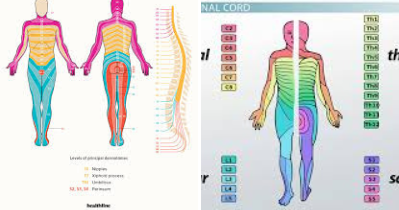
When you look at a dermatomal map, you will see that the lumbar dermatomes are arranged in a somewhat band-like pattern across the lower body. The L1 dermatome covers the area around the inguinal ligament and the upper portion of the front of the thigh. The L2 dermatome extends down the anterior thigh, while the L3 dermatome covers the lower anterior thigh and the knee region. The L4 dermatome is particularly important because it covers the medial aspect of the leg and the big toe, and the L5 dermatome covers the lateral aspect of the leg, the dorsum of the foot, and the other toes. Finally, the S1 dermatome, which is technically a sacral nerve root, covers the lateral foot and the heel, but because it is so frequently implicated in lumbar spine pathology, it is often discussed alongside the lumbar dermatomes. This progression from the groin down to the toes follows a remarkably consistent pattern, but it is essential to remember that everyone is slightly different, and the exact boundaries between dermatomes can vary from person to person.
The Clinical Significance of Dermatomal Patterns
The clinical significance of lumbar dermatomes cannot be overstated, particularly when it comes to diagnosing conditions like radiculopathy, herniated discs, and spinal stenosis. When a patient presents with radiating leg pain, often described as sciatica, the distribution of that pain is one of the most important clues a clinician can use to identify which nerve root is affected. For instance, if a patient has pain that travels down the back of their thigh, around the lateral aspect of the calf, and into the lateral foot, that is a classic pattern for an S1 radiculopathy, which is typically caused by a herniated disc at the L5-S1 level. On the other hand, if the pain travels down the lateral thigh, crosses the front of the shin, and ends up on the top of the foot and the great toe, that points toward an L5 radiculopathy, often resulting from a disc herniation at the L4-L5 level. These clinical patterns are so reliable that they form the backbone of the neurological examination for patients with low back pain.
What makes lumbar dermatomes particularly fascinating is the way they can help clinicians distinguish between different types of pathology. For example, not all leg pain is radicular; sometimes it is referred pain from the sacroiliac joint, the hip, or even the muscles themselves. However, when the pain follows a clear dermatomal distribution, it strongly suggests that a nerve root is being irritated or compressed. This distinction is crucial because the treatment for radicular pain is often different from the treatment for mechanical back pain or joint pathology. A patient with a clear dermatomal pattern of symptoms might be a candidate for epidural steroid injections, surgical decompression, or specific physical therapy interventions designed to relieve pressure on the affected nerve root. Without an understanding of lumbar dermatomes, clinicians would be flying blind, trying to treat symptoms without understanding their underlying cause.
Beyond the initial diagnosis, lumbar dermatomes are also invaluable for monitoring disease progression and treatment response. If a patient undergoes surgery for a herniated disc, the resolution of their dermatomal symptoms is one of the key indicators of surgical success. Similarly, if a patient is being treated conservatively with physical therapy and anti-inflammatory medications, the shrinking of their dermatomal pain pattern can be a reassuring sign that the nerve root is recovering. On the flip side, if the dermatomal symptoms are expanding or becoming more pronounced, that might be a red flag indicating that the nerve compression is worsening and that more aggressive intervention is needed. This dynamic use of dermatomal mapping is part of what makes neurology such a precise and satisfying field; it is all about connecting the dots between anatomy, pathology, and clinical presentation.
Lumbar Radiculopathy and Sciatica
When people talk about lumbar dermatomes, the conversation almost inevitably turns to lumbar radiculopathy and sciatica, two of the most common and debilitating conditions that affect the lower spine. Lumbar radiculopathy refers to the compression or irritation of a nerve root in the lumbar spine, which then leads to pain, numbness, or weakness that radiates along the path of that nerve. Sciatica, on the other hand, is a specific type of radiculopathy that involves the sciatic nerve, which is formed from the L4, L5, S1, S2, and S3 nerve roots. Because the sciatic nerve is such a large and important structure, sciatica can produce symptoms that mimic multiple dermatomal patterns simultaneously, which is why it can sometimes be tricky to diagnose. However, by carefully mapping out the patient’s symptoms onto a dermatomal chart, a skilled clinician can often determine which specific nerve root is the primary culprit.
The most common cause of lumbar radiculopathy is a herniated intervertebral disc, which occurs when the soft, jelly-like center of the disc bulges out through a tear in the tougher outer ring. This bulging material can then press directly on an adjacent nerve root, causing inflammation, pain, and sometimes even neurological deficits. The L4-L5 and L5-S1 levels are the most frequently affected because these levels endure the greatest mechanical stress and undergo the most movement. A herniated disc at the L4-L5 level typically affects the L5 nerve root, producing symptoms in the L5 dermatome, which includes the lateral thigh, the anterior shin, and the dorsum of the foot. A herniated disc at the L5-S1 level affects the S1 nerve root, producing symptoms in the S1 dermatome, which includes the posterior thigh, the calf, and the lateral foot. These patterns are so well-recognized that they are often taught to medical students as classic clinical vignettes.
Another common cause of lumbar radiculopathy is spinal stenosis, which is a narrowing of the spinal canal that can compress multiple nerve roots at once. Unlike a herniated disc, which tends to affect a single nerve root on one side, spinal stenosis often produces bilateral symptoms and can affect multiple dermatomes simultaneously. This is why patients with spinal stenosis often present with a more diffuse pattern of leg pain, numbness, and weakness that can be difficult to map onto a single dermatome. However, even in these cases, understanding lumbar dermatomes is essential because it allows clinicians to determine which levels of the spine are most severely affected. For example, if a patient has symptoms in both the L4 and L5 dermatomes, that suggests that the stenosis is affecting the L4 and L5 nerve roots, which would point toward a stenosis at the L3-L4 or L4-L5 level. This information is critical for planning surgical interventions like laminectomies or decompressive procedures.
Diagnostic Approaches to Dermatomal Assessment
The clinical assessment of lumbar dermatomes begins with a thorough history and physical examination, and it is one of the most rewarding aspects of neurological practice because it allows you to piece together a puzzle using nothing more than your hands, your eyes, and your knowledge of anatomy. During the physical examination, a clinician will typically test each dermatome by assessing sensation in the corresponding skin area. This is usually done using light touch, pinprick, and sometimes temperature discrimination, depending on the specific clinical context. The patient is asked to close their eyes and indicate when they feel the stimulus, and the clinician then compares the sensation on the left side to the right side and also compares different dermatomal areas to each other. This comparative approach is crucial because it helps to identify subtle asymmetries that might not be apparent if you were just testing one area in isolation.
In addition to sensory testing, the neurological examination for Lumbar Dermatomes radiculopathy also includes motor testing and reflex testing, which provide complementary information about nerve root function. For the L4 nerve root, the key muscle to test is the quadriceps, which is responsible for knee extension, and the key reflex is the patellar tendon reflex. For the L5 nerve root, the key muscle is the tibialis anterior, which controls ankle dorsiflexion, and the key reflex is the medial hamstring reflex. For the S1 nerve root, the key muscle is the gastrocnemius-soleus complex, which controls ankle plantarflexion, and the key reflex is the Achilles tendon reflex. By combining sensory findings from the dermatomal examination with motor and reflex findings, a clinician can build a comprehensive picture of which nerve roots are involved and how severely they are affected. This holistic approach is far more powerful than any single test in isolation.
When the clinical examination suggests a specific dermatomal pattern of involvement, the next step is typically to confirm the diagnosis with imaging studies, most commonly magnetic resonance imaging of the Lumbar Dermatomes spine. An MRI can provide exquisitely detailed images of the intervertebral discs, the nerve roots, and the spinal canal, allowing clinicians to visualize exactly what is causing the nerve root compression. In many cases, the MRI findings will correlate beautifully with the dermatomal pattern identified on physical examination, which provides strong confirmation of the diagnosis. However, it is important to note that MRI findings do not always correlate with symptoms; many people have disc bulges or other abnormalities on MRI that are completely asymptomatic. This is why the clinical examination remains the gold standard for diagnosing radiculopathy; the imaging is just there to confirm what the physical examination has already suggested.
Electrodiagnostic Testing and Lumbar Dermatomes
When the clinical picture is unclear or when there is a need to quantify the severity of nerve root injury, electrodiagnostic testing can provide invaluable information. Two primary electrodiagnostic modalities are used in the evaluation of Lumbar Dermatomes radiculopathy: nerve conduction studies and electromyography. Nerve conduction studies involve stimulating a peripheral nerve with a small electrical pulse and measuring the time it takes for the resulting electrical signal to travel along the nerve. This can help to identify whether there is a conduction block or slowing, which might indicate nerve compression. However, nerve conduction studies have limited utility in radiculopathy because the nerve roots themselves are deep within the spine and cannot be directly stimulated. This is where electromyography comes in, as it involves inserting a fine needle electrode into various muscles and listening to the electrical activity that occurs both at rest and during contraction.
Electromyography is particularly useful because it can detect signs of denervation in muscles that are supplied by specific nerve roots. When a nerve root is damaged, the muscles it innervates will often show abnormal electrical activity, such as fibrillation potentials or positive sharp waves, which indicate that the muscle fibers are denervated and are spontaneously firing. By testing multiple muscles in the leg, an electromyographer can often determine which nerve root is affected based on the pattern of muscle involvement. For example, if the tibialis anterior and the extensor hallucis longus are both showing denervation changes, that suggests an L5 radiculopathy, because both of these muscles are primarily innervated by the L5 nerve root. If the gastrocnemius and the soleus are affected, that points toward an S1 radiculopathy. This electrodiagnostic information can be crucial when the clinical examination is equivocal or when there is a need to differentiate between radiculopathy and peripheral neuropathy.
It is worth noting that electrodiagnostic testing has its limitations and is not always necessary for diagnosing Lumbar Dermatomes radiculopathy. In many cases, a careful history and physical examination are sufficient to make the diagnosis and guide treatment. Additionally, electrodiagnostic testing can be uncomfortable for the patient, as it involves electrical stimulation and needle insertion, so it is generally reserved for cases where the diagnosis is uncertain or where there is a need to document the severity of the nerve injury for medicolegal or prognostic purposes. Nevertheless, when used appropriately, electrodiagnostic testing can provide a level of precision that is simply not achievable with clinical examination alone, and it remains a valuable tool in the neurologist’s armamentarium.
Differential Diagnosis of Lumbar Dermatomes Symptoms
One of the most important skills in clinical medicine is the ability to generate a differential diagnosis, which is a list of possible explanations for a patient’s symptoms. When a patient presents with symptoms that fall into a lumbar dermatomal pattern, the differential diagnosis is surprisingly broad and includes not only spinal pathology but also a variety of other conditions that can mimic radiculopathy. Peripheral neuropathies, for example, can produce sensory symptoms that are similar to Lumbar Dermatomes, but they typically affect multiple distal nerve territories rather than a single dermatome. Diabetes, alcohol abuse, and various vitamin deficiencies can all cause peripheral neuropathy, and it is important to consider these possibilities, especially in patients who have risk factors for these conditions. A careful history and physical examination, including a detailed review of the patient’s medical history and medication list, can often help to differentiate radiculopathy from peripheral neuropathy.
Another important condition to consider in the differential diagnosis is meralgia paresthetica, which is a compression of the lateral femoral cutaneous nerve as it passes through the inguinal ligament. This condition produces numbness, tingling, and sometimes burning pain on the lateral aspect of the thigh, which can easily be mistaken for an L2 or L3 radiculopathy. However, there are some key differences; meralgia paresthetica typically does not cause any motor weakness or reflex changes, and the symptoms are purely sensory. Additionally, the symptoms of meralgia paresthetica are often exacerbated by tight clothing, obesity, or prolonged standing, whereas radiculopathy symptoms are typically exacerbated by movements that increase intradiscal pressure, such as coughing, sneezing, or bending forward. Recognizing these subtle differences is essential for making an accurate diagnosis and avoiding unnecessary spinal imaging or surgical interventions.
Hip pathology is another common mimicker of Lumbar Dermatomes, particularly osteoarthritis of the hip or avascular necrosis of the femoral head. Patients with hip pathology often present with pain in the groin, the anterior thigh, or the buttock, which can easily be confused with L2 or L3 radiculopathy. However, hip pathology typically causes pain that is worsened by weight-bearing activities like walking or standing, whereas radiculopathy pain is often worse with sitting or bending. Additionally, hip pathology usually does not cause any sensory changes or reflex abnormalities, which helps to differentiate it from radiculopathy. A thorough physical examination, including assessment of hip range of motion and provocative maneuvers like the Patrick test, can help to identify hip pathology and prevent misdiagnosis. This is a perfect example of why clinical acumen and a detailed understanding of anatomy are so important; the key to making an accurate diagnosis is often in the details.
Treatment Approaches for Lumbar Dermatomes Radiculopathy
The treatment of Lumbar Dermatomes radiculopathy can be broadly divided into conservative management, interventional procedures, and surgical interventions, and the choice of treatment depends on a variety of factors, including the severity of the symptoms, the degree of neurological deficit, and the patient’s overall health and preferences. Conservative management is typically the first-line approach and includes a combination of physical therapy, pain medications, and lifestyle modifications. Physical therapy for lumbar radiculopathy focuses on strengthening the core muscles, improving flexibility, and correcting postural imbalances that might be contributing to nerve root irritation. Pain medications, including nonsteroidal anti-inflammatory drugs, muscle relaxants, and sometimes neuropathic pain medications like gabapentin or pregabalin, can help to manage symptoms while the patient is undergoing physical therapy. Lifestyle modifications, such as weight loss, smoking cessation, and ergonomic adjustments, are also important for reducing mechanical stress on the lumbar spine and preventing recurrence.
When conservative management is not sufficient to control symptoms, interventional procedures may be considered. Epidural steroid injections are one of the most commonly performed interventional procedures for Lumbar Dermatomes radiculopathy and involve injecting a corticosteroid medication into the epidural space around the affected nerve root. The steroid works by reducing inflammation and swelling, which can relieve pressure on the nerve root and provide significant pain relief. These injections can be performed with or without fluoroscopic guidance, but most pain specialists prefer to use fluoroscopy to ensure accurate placement of the needle. Epidural steroid injections are not a cure for radiculopathy; they are a temporizing measure that can provide relief for weeks to months, allowing the patient to participate more effectively in physical therapy and other rehabilitative interventions. In some cases, a series of injections may be needed to achieve adequate symptom control.
Surgical intervention is generally reserved for patients who have failed conservative management or who have significant neurological deficits, such as progressive weakness, bowel or bladder dysfunction, or intractable pain that is unresponsive to all other treatments. The most common surgical procedure for Lumbar Dermatomes radiculopathy is a discectomy, which involves removing the portion of the herniated disc that is compressing the nerve root. This can be performed through a variety of approaches, including open surgery, microdiscectomy, and endoscopic discectomy, and the choice of approach depends on the surgeon’s preference and the specific characteristics of the herniated disc. In patients with spinal stenosis, a decompressive laminectomy may be performed, which involves removing the lamina and other bony structures to create more space for the nerve roots. Surgical outcomes for lumbar radiculopathy are generally excellent, with most patients experiencing significant improvement in their symptoms, particularly if the surgery is performed before permanent nerve damage has occurred.
Rehabilitation and Long-Term Management
Rehabilitation is a critical component of the overall management plan for patients with Lumbar Dermatomes radiculopathy, and it should begin as soon as the acute phase of the condition has resolved. The goals of rehabilitation are to restore normal function, prevent recurrence, and empower the patient to take an active role in their own recovery. A comprehensive rehabilitation program for lumbar radiculopathy typically includes a combination of therapeutic exercises, manual therapy, and patient education. Therapeutic exercises are designed to strengthen the muscles that support the lumbar spine, improve flexibility, and correct movement patterns that might be contributing to nerve root irritation. Specific exercises might include core stabilization exercises, stretching of the hamstrings and hip flexors, and strengthening of the gluteal muscles. Manual therapy techniques, such as joint mobilization and soft tissue massage, can help to reduce pain, improve mobility, and facilitate the healing process.
Patient education is another crucial component of rehabilitation, as it empowers patients to understand their condition and take steps to prevent recurrence. Education typically covers topics such as proper body mechanics, ergonomic principles, and strategies for managing activities of daily living. Patients are taught how to lift objects safely, how to sit and stand with proper posture, and how to modify their activities to avoid exacerbating their symptoms. They are also educated about the importance of maintaining a healthy weight, staying physically active, and avoiding prolonged periods of sitting or standing. This educational component is often the most valuable part of rehabilitation because it gives patients the tools they need to manage their condition effectively over the long term. Without this knowledge, patients are at high risk of recurrence, as they may inadvertently engage in activities that put excessive stress on their lumbar spine.
Long-term management of lumbar radiculopathy involves a commitment to ongoing self-care and regular follow-up with healthcare providers. Patients who have had a radiculopathy are at increased risk for future episodes, so they need to remain vigilant about their spinal health. This might involve continuing with a home exercise program, periodically checking in with a physical therapist or personal trainer, and scheduling regular appointments with their primary care physician or spine specialist. For patients who have undergone surgery, regular follow-up is particularly important to monitor for any signs of recurrence or complications. It is also important for patients to be aware of red flag symptoms, such as new or worsening weakness, bowel or bladder dysfunction, or saddle anesthesia, which could indicate a more serious condition like cauda equina syndrome. By staying proactive about their spinal health, patients can minimize their risk of recurrence and maintain a high quality of life.
The Role of Imaging in Dermatomal Diagnosis
Magnetic resonance imaging is the imaging modality of choice for evaluating lumbar radiculopathy because it provides superior soft tissue contrast and can visualize the intervertebral discs, the nerve roots, and the spinal canal with remarkable detail. An MRI of the lumbar spine can identify disc herniations, spinal stenosis, foraminal stenosis, and other structural abnormalities that might be compressing nerve roots. The MRI findings can then be correlated with the patient’s clinical presentation, including the dermatomal pattern of symptoms, to confirm the diagnosis and plan treatment. In many cases, the MRI will show a disc herniation at the level that corresponds to the patient’s dermatomal symptoms, which provides strong support for the diagnosis. However, it is important to remember that MRI findings do not always correlate with symptoms, and many asymptomatic individuals have abnormal findings on MRI. This is why the clinical examination remains the cornerstone of diagnosis, with imaging serving as a confirmatory tool.
Computed tomography and myelography are other imaging modalities that can be used in the evaluation of lumbar radiculopathy, although they are less commonly used than MRI. CT is particularly useful for evaluating bony abnormalities, such as facet joint hypertrophy or osteophyte formation, which can contribute to foraminal stenosis. Myelography involves injecting a contrast dye into the subarachnoid space and then taking X-rays or CT images to visualize the spinal cord and nerve roots. This can be useful in patients who cannot undergo MRI, such as those with pacemakers or other metallic implants. However, myelography is an invasive procedure that carries a risk of complications, such as headaches, infection, or nerve injury, so it is generally reserved for cases where MRI is contraindicated or where additional information is needed. In clinical practice, MRI is overwhelmingly the preferred imaging modality for evaluating lumbar radiculopathy, and it is the standard of care in most settings.
It is also worth noting that imaging findings must be interpreted in the context of the patient’s clinical presentation, and not all disc herniations or stenotic changes are clinically significant. Asymptomatic disc herniations are surprisingly common, with studies showing that up to 30 percent of asymptomatic individuals have disc bulges or herniations on MRI. This is why it is so important to correlate imaging findings with the patient’s symptoms and physical examination findings. A disc herniation that is not causing any symptoms does not require treatment, even if it looks impressive on the MRI. Conversely, a patient with classic radicular symptoms may have only a small disc herniation on MRI, but that small herniation might be in exactly the right location to compress the nerve root. This is the art of clinical medicine: synthesizing information from multiple sources to arrive at the correct diagnosis and treatment plan.
Special Considerations for Cauda Equina Syndrome
Cauda equina syndrome is a rare but serious condition that requires prompt recognition and treatment, and it is one of the few true emergencies in spine medicine. The cauda equina, which translates to horse’s tail, is a bundle of nerve roots that extends from the bottom of the spinal cord down through the lumbar and sacral spine. When this bundle is compressed, typically by a large central disc herniation or by a mass lesion, it can cause a constellation of symptoms that includes bilateral leg pain, weakness, and sensory changes, as well as bowel and bladder dysfunction and saddle anesthesia. Saddle anesthesia refers to numbness or tingling in the perineal area, which would be supplied by the S2 through S4 dermatomes. The presence of saddle anesthesia, along with bowel or bladder dysfunction, is a red flag that should prompt immediate evaluation and possible surgical intervention.
The diagnosis of cauda equina syndrome is primarily clinical, and it is one of those diagnoses that you cannot afford to miss. Any patient who presents with new-onset bowel or bladder dysfunction, bilateral leg symptoms, or saddle anesthesia should be evaluated emergently, with MRI being the imaging modality of choice to identify the underlying cause of the compression. The treatment for cauda equina syndrome is surgical decompression, and the timing of surgery is critical; outcomes are significantly better when surgery is performed within 24 to 48 hours of symptom onset, and delays can lead to permanent neurological deficits. This is why it is so important to maintain a high index of suspicion for cauda equina syndrome in any patient who presents with acute low back pain and radicular symptoms. A thorough history and physical examination, including a detailed assessment of bowel and bladder function and perineal sensation, is essential for identifying this condition and initiating appropriate treatment.
Conclusion: Integrating Dermatomal Knowledge into Practice
Understanding lumbar dermatomes is not just an academic exercise; it is a practical skill that has direct implications for patient care. Whether you are a physical therapist designing a rehabilitation program, a primary care physician managing a patient with low back pain, or a spine surgeon planning an operation, your knowledge of lumbar dermatomes will guide your decision-making and improve your patient outcomes. By being able to map a patient’s symptoms onto a dermatomal chart, you can identify the likely nerve root involved, determine the level of the spine that is causing the problem, and tailor your treatment accordingly. This precision is what distinguishes excellent clinical care from mediocre care, and it is one of the reasons why a solid foundation in anatomy and neurology is so important for all healthcare providers.
As our understanding of the nervous system continues to evolve, so too does our appreciation for the complexity and significance of lumbar dermatomes. Advances in imaging technology, electrodiagnostic testing, and surgical techniques have all enhanced our ability to diagnose and treat lumbar radiculopathy, but the basic principles of dermatomal mapping remain as relevant today as they were a century ago. The dermatome is a testament to the incredible organization of the human nervous system, and it serves as a constant reminder that the body is a beautifully integrated system where structure and function are intimately linked. For the patient who is struggling with unexplained leg pain, the dermatome can provide an answer, a direction, and ultimately, a path to recovery. For the clinician, the dermatome is a powerful tool that bridges the gap between anatomy and clinical medicine, transforming abstract knowledge into tangible benefits for the patient. So the next time you find yourself tracing the distribution of a patient’s leg pain, take a moment to appreciate the rich history and profound clinical significance of the humble lumbar dermatome.
The Fossa Ovalis: A Comprehensive Exploration of the Heart’s Hidden Landmark