
The human heart is an incredible organ, a tireless pump that works day and night to keep us Fossa Ovalis. Within its complex structure lies a fascinating feature that many people have never heard of, yet it plays a crucial role in our development and continues to intrigue medical professionals worldwide. This feature is the fossa ovalis, a small but significant depression found in the wall between the heart’s upper chambers. While it might seem like just another anatomical detail, this structure tells a remarkable story about how our circulatory system evolves from the moment we are conceived until the day we are born.
The fossa ovalis is not just a random indentation; it represents a remnant of our fetal existence, a scar that marks the closure of a vital passageway that once allowed blood to bypass the lungs. Understanding this structure is essential for anyone interested in cardiology, anatomy, or even just the miraculous ways our bodies adapt to life outside the womb. In this comprehensive article, we will explore every aspect of the fossa ovalis, from its embryonic origins to its clinical significance in modern medicine, and uncover why this tiny depression deserves far more attention than it typically receives.
The Embryological Journey of the Fossa Ovalis
The story of the fossa ovalis begins long before a baby takes its first breath, during the intricate process of fetal development. In the early weeks of gestation, the heart forms from a simple tube that eventually folds and partitions into four distinct chambers. During this period, the fetal circulatory system operates very differently from that of a newborn, primarily because the lungs are not yet functional. Instead of receiving oxygen from the air, the developing baby relies entirely on the placenta for oxygenated blood, which means the heart must be cleverly designed to direct blood flow away from the lungs and toward the rest of the body.
This is where the foramen ovale enters the picture. The foramen ovale is essentially an opening in the interatrial septum, the wall that separates the left and right atria. This opening allows oxygen-rich blood coming from the placenta through the inferior vena cava to pass directly from the right atrium into the left atrium, thereby bypassing the non-functional fetal lungs. This clever adaptation ensures that the developing brain and other vital organs receive the most highly oxygenated blood available, which is crucial for proper growth and development during the nine months of pregnancy.
As the fetal heart grows and matures, the foramen ovale remains patent, or open, throughout gestation, maintained by a delicate balance of pressures between the two atria. The right atrial pressure is slightly higher than the left, which keeps the flap-like valve of the foramen ovale pushed open, allowing blood to flow freely from right to left. This physiological shunt is one of the most remarkable examples of how the human body adapts to different environments, and it functions flawlessly for the entire duration of the pregnancy, ensuring that the fetus receives all the oxygen and nutrients it needs to thrive.
However, the moment of birth changes everything. When a baby takes its first breath, the lungs expand, and pulmonary vascular resistance drops dramatically, causing a sudden increase in blood flow to the lungs. At the same time, the pressure in the left atrium rises as more blood returns from the newly functional lungs. This reversal of pressure gradients causes the valve of the foramen ovale to close against the interatrial septum, effectively sealing the opening. Over the following months, this closure becomes permanent as the tissues fuse together, leaving behind only a shallow depression in the atrial septum, which we now call the fossa ovalis.
The transition from foramen ovale to fossa ovalis is not always instantaneous or complete. In some cases, the closure may take longer, and in approximately twenty to thirty percent of the population, the foramen ovale never fully seals, leaving a small flap-like opening that can persist into adulthood. This persistent opening is known as a patent foramen ovale, or PFO, and while it often causes no symptoms, it can occasionally be associated with certain medical conditions. The fossa ovalis itself, however, represents the successful closure of this fetal shunt in the majority of individuals, marking the completion of one of the most important physiological transitions we will ever experience.
Anatomical Structure and Location of the Fossa Ovalis
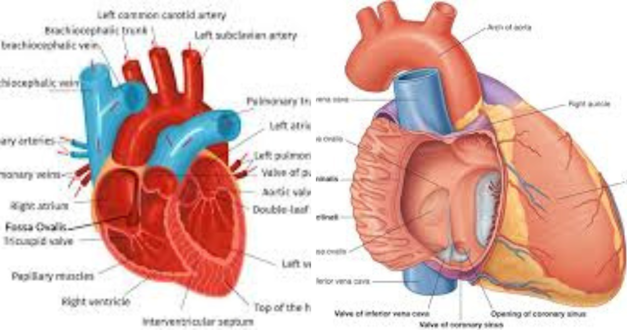
The fossa ovalis is situated in the interatrial septum, which is the muscular wall that separates the left and right atria of the heart. More specifically, it is located in the lower portion of this septum, just above the opening of the inferior vena cava and slightly posterior to the limbus, which is the prominent ridge that surrounds it. This precise location is clinically significant because it serves as a landmark for various cardiac procedures, and understanding its exact position is essential for interventional cardiologists and cardiac surgeons alike.
In terms of its physical appearance, the fossa ovalis resembles a shallow, oval-shaped depression or dimple in the atrial septum. Its dimensions can vary considerably from person to person, but on average, it measures approximately two centimeters in length and one centimeter in width. The floor of the fossa ovalis is typically thin and membranous, composed of a remnant of the septum primum, which is one of the embryonic structures that participated in the formation of the interatrial septum. This thinness is clinically relevant because it makes the fossa ovalis a common site for iatrogenic perforations during cardiac catheterization procedures, and it also explains why transseptal punctures are often performed at this location.
Surrounding the fossa ovalis is a thick, crescent-shaped muscular ridge known as the limbus of the fossa ovalis, or the annulus ovalis. This prominent border is formed by the septum secundum, another embryonic structure that grows alongside the septum primum during development. The limbus is most prominent on the superior and anterior margins of the fossa ovalis, giving the structure its characteristic appearance of a depression surrounded by a raised rim. This anatomical relationship is often compared to a crater or a dimple, with the limbus serving as the elevated outer edge and the fossa itself forming the depressed center.
The relationship between the fossa ovalis and surrounding structures is crucial for understanding its clinical significance. It lies in close proximity to the atrioventricular node, the bundle of His, and the coronary sinus, all of which are vital components of the heart’s conduction system. This proximity means that any procedure performed in this area, such as transseptal puncture or device closure of a patent foramen ovale, carries a risk of damaging these important structures, which could potentially lead to heart block or other conduction abnormalities. Therefore, interventional cardiologists must have an intimate knowledge of the anatomy of the fossa ovalis and its surrounding structures to perform these procedures safely and effectively.
From a histological perspective, the fossa ovalis is composed of a unique type of tissue that reflects its embryonic origins. The floor of the fossa is made of a thin layer of connective tissue covered by endothelium on both the right and left atrial surfaces. This tissue is relatively acellular and lacks the dense muscularity of the surrounding interatrial septum, which is why it is often more susceptible to perforation or tearing. The limbus, on the other hand, is composed of dense muscular tissue that provides structural support and helps maintain the integrity of the septum. Understanding these histological differences is essential for pathologists and researchers who study the heart’s structure and its response to various diseases and interventions.
The Role of the Fossa Ovalis in Fetal Circulation
The fossa ovalis represents a vestige of a system that was absolutely essential for fetal survival, a testament to the remarkable adaptability of the developing cardiovascular system. During fetal life, the presence of a patent foramen ovale is not just normal but absolutely necessary, as it allows blood to be diverted away from the lungs, which are non-functional and filled with fluid rather than air. This diversion is part of a larger system of shunts that exist in the fetal heart, including the ductus arteriosus and the ductus venosus, all working together to ensure that the developing baby receives the best possible oxygen supply.
The circulation of blood through the fetal heart follows a very specific pathway that prioritizes the delivery of oxygenated blood to the brain and other vital organs. Blood returning from the placenta through the umbilical vein is rich in oxygen and nutrients, and much of this blood bypasses the liver through the ductus venosus before entering the inferior vena cava. From the inferior vena cava, this oxygenated blood flows into the right atrium, where the majority of it is directed through the foramen ovale into the left atrium, thanks to the presence of a structure called the crista dividens. This preferential streaming ensures that the most oxygenated blood is delivered to the left side of the heart, from which it is pumped through the aorta to supply the brain and the rest of the upper body.
The rest of the blood entering the right atrium is mixed with deoxygenated blood returning from the superior vena cava, and this mixture is then directed into the right ventricle and pumped out through the pulmonary artery. However, because the fetal lungs are not yet ventilated, the pulmonary vascular resistance is high, and most of this blood bypasses the lungs through the ductus arteriosus, a vessel that connects the pulmonary artery to the aorta. This intricate system of shunts ensures that the developing fetus has a continuous supply of oxygenated blood for growth and development, while simultaneously preparing the cardiovascular system for the dramatic changes that will occur at birth.
The mechanisms that maintain the patency of the foramen ovale during fetal life are complex and involve a delicate interplay of pressure gradients, hormonal signals, and anatomical factors. The primary driving force is the pressure difference between the two atria, with right atrial pressure being slightly higher than left atrial pressure throughout most of gestation. This pressure gradient is maintained by several factors, including the high resistance of the fetal pulmonary vasculature, which keeps right ventricular pressure elevated, and the relatively low resistance of the systemic circulation, which keeps left atrial pressure lower. In addition, the valve of the foramen ovale, which is a flap-like structure formed by the septum primum, is held open by the flow of blood from the inferior vena cava, which directs oxygenated blood directly against this valve.
Hormonal factors also play a role in maintaining the patency of the foramen ovale during fetal development. Prostaglandins, in particular, are known to play a role in keeping both the foramen ovale and the ductus arteriosus open throughout gestation. These hormonally mediated mechanisms ensure that the fetal shunts remain functional even as the heart grows and develops, and they represent yet another layer of regulation that protects the developing fetus from the consequences of premature closure of these essential passages. Understanding these hormonal influences is crucial for neonatologists and obstetricians who manage pregnancies complicated by conditions that might affect fetal circulation.
Transition at Birth: Closure of the Foramen Ovale
The moment of birth is perhaps the most dramatic event in human life, and no organ undergoes more profound changes in such a short time than the heart. The transition from fetal to neonatal circulation requires a series of carefully orchestrated events, and the closure of the foramen ovale is one of the most important milestones in this process. This transformation begins with the baby’s first breath, which triggers a cascade of physiological changes that ultimately result in the permanent closure of the foramen ovale and the formation of the fossa ovalis.
When a newborn takes its first breath, the lungs expand, and the pulmonary vascular resistance drops dramatically as the pulmonary vessels dilate in response to increased oxygenation. This sudden decrease in resistance allows blood to flow more freely through the pulmonary circulation, leading to a significant increase in pulmonary venous return to the left atrium. At the same time, the clamping of the umbilical cord removes the placental circulation from the equation, which causes a reduction in venous return to the right atrium and a subsequent drop in right atrial pressure. The net effect of these changes is that left atrial pressure now exceeds right atrial pressure, causing the valve of the foramen ovale to be pushed against the interatrial septum and effectively sealing the opening.
This functional closure of the foramen ovale occurs within minutes to hours after birth and is initially reversible if right atrial pressure were to rise again due to certain medical conditions. However, over the ensuing weeks and months, the tissues of the septum primum and septum secundum undergo fusion, forming a permanent seal that is known as the fossa ovalis. This anatomical closure is typically complete by around the age of two years, although in many individuals, a small remnant of the opening may persist, resulting in a probe-patent foramen ovale that can be opened under certain physiological conditions.
The process of permanent closure involves several cellular and molecular mechanisms. The valve of the foramen ovale, which is composed of the septum primum, fuses with the muscular tissue of the septum secundum through a process of cellular proliferation and extracellular matrix remodeling. This fusion is mediated by various growth factors and cytokines that promote tissue adhesion and healing, creating a robust seal that can withstand the pressures and stresses of postnatal life. In some cases, however, this fusion is incomplete, leading to a persistent flap-like opening that can allow blood to pass between the atria under certain conditions, such as during coughing, straining, or other activities that increase right atrial pressure.
It is important to note that while the foramen ovale closes functionally in the vast majority of newborns, a significant proportion of the population retains a patent foramen ovale throughout their lives. Studies have shown that a patent foramen ovale is present in approximately twenty to thirty percent of adults, although the incidence decreases with age as the tissues continue to fuse over time. The presence of a patent foramen ovale is generally considered a benign anatomical variant, but it has been associated with certain medical conditions, particularly in younger patients, which we will explore in more detail later in this article.
Clinical Significance and Conditions Associated with the Fossa Ovalis
The fossa ovalis, despite its small size and relatively simple structure, has significant clinical implications across a wide range of medical conditions. Its location in the interatrial septum makes it a critical landmark for several interventional procedures, and its embryological origins mean that abnormalities in its formation or closure can lead to a variety of congenital and acquired conditions. Understanding the clinical significance of the fossa ovalis is essential for cardiologists, cardiac surgeons, and general practitioners alike, as it can affect everything from diagnostic imaging to surgical planning.
One of the most important clinical associations involving the fossa ovalis is the patent foramen ovale, which we touched upon earlier. A patent foramen ovale occurs when the foramen ovale fails to close completely after birth, leaving a small flap-like opening between the atria that can allow blood to shunt from right to left under certain conditions. This right-to-left shunt can allow small blood clots, air bubbles, or other emboli to bypass the pulmonary circulation and enter the systemic circulation, potentially leading to a condition known as paradoxical embolism. If these emboli travel to the brain, they can cause a cryptogenic stroke, which is a stroke with no obvious cause, and this association has been the subject of extensive research and clinical debate in recent years.
The relationship between patent foramen ovale and cryptogenic stroke is complex and not fully understood, but it has led to the development of device closure procedures that aim to seal the opening and prevent future embolic events. These procedures involve the insertion of a specialized device through the fossa ovalis, which is then expanded to close the opening permanently. While the clinical benefit of this procedure remains a matter of ongoing research and debate, it has become increasingly common in selected patients, particularly those who have experienced a cryptogenic stroke and have a large or significant patent foramen ovale. The success of these procedures depends on a thorough understanding of the anatomy of the fossa ovalis and the surrounding structures.
Another condition associated with the fossa ovalis is atrial septal aneurysm, which is a congenital abnormality characterized by a bulging of the interatrial septum, often involving the region of the fossa ovalis. This bulging can be present at birth or develop later in life, and it is frequently associated with a patent foramen ovale. Atrial septal aneurysms have been identified as an independent risk factor for cryptogenic stroke, likely because they can promote the formation of blood clots within the aneurysm or facilitate the shunting of emboli through a patent foramen ovale. The diagnosis and management of atrial septal aneurysms require careful imaging and often involve the same considerations as those for patent foramen ovale.
The fossa ovalis also plays a role in several congenital heart defects that affect the interatrial septum. Secundum atrial septal defects, which are openings in the interatrial septum that are located in the region of the fossa ovalis, are among the most common congenital heart defects in adults. These defects occur when the septum primum and septum secundum fail to fuse completely, leaving a hole that allows blood to flow between the atria. The size and location of the defect can vary considerably, and treatment options range from conservative management to surgical or device closure, depending on the severity of the symptoms and the size of the defect.
Cardiac tumors, although rare, can also affect the fossa ovalis. Myxomas, which are the most common primary cardiac tumors, frequently arise from the interatrial septum in the region of the fossa ovalis. These benign tumors can cause a range of symptoms, including embolic events, obstruction of blood flow, and arrhythmias, and they require surgical removal for definitive treatment. The location of myxomas in the fossa ovalis is thought to be related to the embryological origins of this structure, as the residual embryonic tissues in this area may be more prone to tumor formation.
Diagnostic Imaging and the Fossa Ovalis
Advances in medical imaging have dramatically improved our ability to visualize the fossa ovalis and assess its structure and function. Echocardiography, particularly transesophageal echocardiography, is the primary imaging modality used to evaluate the interatrial septum and identify conditions such as patent foramen ovale, atrial septal defects, and atrial septal aneurysms. The fossa ovalis can be clearly seen on these imaging studies as a thin depression in the interatrial septum, and its relationship to surrounding structures can be accurately assessed.
Transthoracic echocardiography is often the first imaging modality used to evaluate the heart in patients with suspected interatrial abnormalities. While this non-invasive technique can often visualize the fossa ovalis and detect gross structural abnormalities, it has limitations in terms of resolution and the ability to accurately assess shunting through a patent foramen ovale. For these reasons, transesophageal echocardiography is frequently employed when a more detailed evaluation is needed, as the proximity of the probe to the heart allows for much higher resolution images of the interatrial septum and the fossa ovalis.
Contrast echocardiography is another valuable tool for evaluating the fossa ovalis and detecting the presence of a patent foramen ovale. This technique involves the injection of agitated saline or a contrast agent into a peripheral vein, followed by echocardiographic visualization of the micro-bubbles as they traverse the cardiac chambers. In the presence of a patent foramen ovale, micro-bubbles can be seen crossing from the right atrium to the left atrium within a few cardiac cycles, confirming the presence of a right-to-left shunt. The sensitivity of contrast echocardiography can be increased by performing a Valsalva maneuver or other actions that increase right atrial pressure, which helps to open the flap of the patent foramen ovale and demonstrate the shunt.
Cardiac magnetic resonance imaging and cardiac computed tomography are also increasingly used to evaluate the fossa ovalis and the interatrial septum. These advanced imaging techniques provide excellent spatial resolution and can be particularly useful in cases where echocardiographic imaging is suboptimal or when additional information about the heart and surrounding structures is needed. Cardiac magnetic resonance imaging, in particular, can provide functional information about blood flow and shunting, as well as detailed anatomical information, making it a valuable tool for the comprehensive evaluation of patients with suspected interatrial abnormalities.
Invasive imaging techniques, such as intracardiac echocardiography, are sometimes used during interventional procedures to guide the placement of closure devices or to perform transseptal punctures. Intracardiac echocardiography involves placing a specialized ultrasound probe directly inside the heart via a catheter, allowing for real-time imaging of the fossa ovalis and surrounding structures. This technique provides excellent image quality and can be used to accurately guide the passage of a needle through the fossa ovalis during transseptal puncture, reducing the risk of complications such as pericardial effusion or puncture of the aortic root.
The Fossa Ovalis and Interventional Procedures
The fossa ovalis has become an increasingly important landmark in interventional cardiology, as it serves as the preferred site for transseptal puncture during various cardiac procedures. Transseptal puncture involves the passage of a specialized needle through the interatrial septum to access the left side of the heart from the right side, allowing for the performance of procedures such as mitral valvuloplasty, left atrial appendage closure, and catheter ablation for atrial fibrillation. The thin, membranous nature of the fossa ovalis makes it ideal for this purpose, as it allows the needle to pass through the septum with relative ease while minimizing the risk of damage to surrounding structures.
The technique of transseptal puncture requires precise understanding of the anatomy of the fossa ovalis and the surrounding structures. The needle is typically advanced from the femoral vein through the inferior vena cava into the right atrium, where it is positioned against the fossa ovalis under fluoroscopic or echocardiographic guidance. The ideal puncture site is at the center of the fossa ovalis, which is typically located in the lower portion of the interatrial septum, just above the opening of the inferior vena cava and slightly posterior to the limbus. Once the needle is advanced through the septum, a guidewire is passed into the left atrium, followed by a sheath that provides access for the therapeutic device.
Complications of transseptal puncture can include pericardial effusion, cardiac tamponade, aortic puncture, and damage to the conduction system. These complications are relatively rare in experienced hands, but they can be catastrophic if they occur, which underscores the importance of a thorough understanding of the anatomy and careful imaging guidance during the procedure. The development of newer imaging techniques, such as three-dimensional transesophageal echocardiography and intracardiac echocardiography, has further improved the safety of transseptal puncture by providing more detailed visualization of the fossa ovalis and its relationship to surrounding structures.
Beyond transseptal puncture, the fossa ovalis is also the site for the placement of devices to close a patent foramen ovale or to treat secundum atrial septal defects. These devices are designed to be deployed through the fossa ovalis and then expanded to seal the opening, effectively closing the communication between the atria. The deployment of these devices requires careful positioning and imaging to ensure that they are properly seated and that they do not interfere with the function of the surrounding structures, such as the atrioventricular valves or the pulmonary veins.
The success of these interventional procedures has revolutionized the management of many cardiac conditions, offering less invasive alternatives to open-heart surgery for many patients. However, the use of these devices is not without controversy, particularly in the case of patent foramen ovale closure for cryptogenic stroke. Ongoing clinical trials are continuing to evaluate the efficacy and safety of these procedures, helping to clarify the optimal patient selection and treatment strategies for various conditions affecting the fossa ovalis.
Evolution of Knowledge and Historical Perspectives
Our understanding of the fossa ovalis has evolved considerably over the centuries, from the early descriptions of ancient anatomists to the sophisticated imaging and interventional techniques available today. The recognition of the foramen ovale and its significance in fetal circulation dates back to the work of ancient physicians, who observed the structures of the fetal heart but lacked the understanding of their physiological significance. It was not until the Renaissance that anatomists such as Leonardo da Vinci and William Harvey began to elucidate the true nature of the heart’s structure and function, paving the way for modern cardiovascular medicine.
William Harvey’s publication of “Exercitatio Anatomica de Motu Cordis et Sanguinis in Animalibus” in 1628 represented a landmark in the understanding of the cardiovascular system, and his description of the circulation of blood helped to clarify the role of the foramen ovale in fetal life. Harvey recognized that the foramen ovale allowed blood to bypass the lungs in the fetus, and he correctly understood that its closure after birth was a critical step in the transition to postnatal circulation. This insight laid the foundation for the study of congenital heart defects and the development of treatments for conditions affecting the interatrial septum.
Throughout the nineteenth and twentieth centuries, advances in anatomy, pathology, and surgical technique continued to expand our understanding of the fossa ovalis and its clinical significance. The development of cardiac catheterization in the mid-twentieth century allowed researchers to study the heart’s structure and function in living patients, providing unprecedented insight into conditions such as patent foramen ovale and atrial septal defects. This era also saw the emergence of cardiac surgery, with pioneering surgeons performing the first successful repairs of congenital heart defects, including those involving the interatrial septum.
The later decades of the twentieth century brought the development of echocardiography, which revolutionized the diagnosis of cardiac conditions and allowed clinicians to visualize the fossa ovalis non-invasively. The introduction of transesophageal echocardiography in the 1980s provided even better visualization of the interatrial septum and paved the way for the development of interventional techniques such as transseptal puncture and device closure of patent foramen ovale and atrial septal defects. These advances have dramatically improved the outcomes for patients with conditions affecting the fossa ovalis, and they continue to evolve with ongoing technological developments and clinical research.
Today, the fossa ovalis is recognized as more than just an anatomical curiosity; it is a crucial structure with significant clinical implications across multiple medical disciplines. The study of this small depression has provided insights into fetal development, congenital heart disease, and the mechanisms of stroke, and it continues to be a focus of active clinical and basic science research. As we continue to develop new imaging and interventional techniques, our understanding of the fossa ovalis will undoubtedly continue to expand, leading to even better outcomes for patients with conditions affecting this fascinating structure.
Fossa Ovalis in Surgical Considerations
The fossa ovalis and the interatrial septum present specific challenges and considerations for cardiac surgeons. When operating on the heart, especially in procedures involving the left atrium or mitral valve, the interatrial septum is a frequently used approach to access these structures. The fossa ovalis, being the thinnest part of the septum, is an ideal location for creating an atriotomy, or a surgical incision, that allows the surgeon to reach the left side of the heart while minimizing damage to the surrounding muscular structures.
In cardiac surgery, the incision through the fossa ovalis is often termed a “septal approach” or “Sondergaard’s groove incision.” When performed correctly, this approach provides excellent exposure to the mitral valve, left atrium, and other left-sided structures. The closure of this surgical incision must be meticulous to prevent residual shunting or the development of an acquired atrial septal defect. Surgeons utilize careful suturing techniques to ensure that the closure is secure and that the normal anatomy of the interatrial septum is preserved.
However, there are important considerations that must be taken into account when operating in this area. The proximity of the atrioventricular node and the bundle of His means that sutures placed too deeply or too posteriorly could disrupt the cardiac conduction system, leading to heart block that might require the placement of a permanent pacemaker. Additionally, the thin nature of the fossa ovalis tissue requires careful handling to prevent tearing or suture dehiscence, which could lead to complications such as bleeding or residual shunting.
In reoperations, the interatrial septum may be scarred, altered, or partially replaced by prosthetic materials from previous surgeries, making the identification of the fossa ovalis more challenging and potentially increasing the risk of complications. Surgeons must be prepared to adapt their approach based on the specific anatomy encountered in each patient, sometimes using alternative routes to access the left atrium when the fossa ovalis is not clearly identifiable or when the interatrial septum has been compromised by prior interventions.
Furthermore, in the context of congenital heart surgery, the fossa ovalis is a critical structure for surgeons repairing atrial septal defects. When closing a secundum defect, which is located in the region of the fossa ovalis, surgeons must differentiate between the true fossa and the defect to ensure that the patch or suture line is placed correctly, preserving a normal interatrial septum and maintaining the appropriate size and function of both atria. This differentiation often requires a combination of visual inspection, palpation, and sometimes intraoperative echocardiographic guidance.
The Fossa Ovalis in Veterinary Medicine
While this article focuses primarily on human anatomy and clinical significance, it is worth briefly exploring the presence and importance of the fossa ovalis in veterinary medicine, particularly in the context of companion animals and comparative anatomy. In domestic animals such as dogs, cats, and horses, the fossa ovalis has a similar embryological origin and anatomical location, representing the site of the former foramen ovale that closed after birth.
In veterinary cardiology, the fossa ovalis can be involved in various congenital and acquired conditions that affect companion animals. Atrial septal defects, including those in the region of the fossa ovalis, occur in dogs and cats, although they are less common than in humans. These defects can cause similar clinical signs, including exercise intolerance, respiratory distress, and in some cases, the development of heart failure. The diagnosis and management of these defects in animals often parallel the approach used in human medicine, with echocardiography being the primary diagnostic tool.
The patent foramen ovale is also recognized in animals, particularly in dogs, where it has been associated with conditions such as pulmonary hypertension and cyanotic heart disease. In veterinary practice, the clinical significance of a patent foramen ovale is often considered in the context of other underlying cardiac or pulmonary conditions. The management of these conditions in animals is generally conservative, although interventional closure techniques are being developed and refined in veterinary medicine.
In addition to its clinical relevance, the study of the fossa ovalis in various animal species has contributed to our understanding of comparative cardiovascular anatomy and evolution. The presence of a foramen ovale and its subsequent closure into a fossa ovalis is a feature of all mammals, and its study across species provides insights into the evolutionary pressures that have shaped the mammalian cardiovascular system. These comparative studies highlight the remarkable conservation of certain anatomical features across species, underscoring the fundamental importance of efficient cardiovascular function for survival and adaptation.
The Future of Fossa Ovalis Research
The fossa ovalis continues to be a subject of active research, with ongoing investigations into its embryonic development, anatomical variations, and clinical significance. Recent advances in genetics, molecular biology, and imaging techniques are providing new insights into the mechanisms that regulate foramen ovale closure and the factors that contribute to its persistence in some individuals. These studies are likely to have important clinical implications, potentially leading to new strategies for preventing and managing conditions associated with the fossa ovalis.
One particularly exciting area of research involves the genetic determinants of patent foramen ovale and atrial septal defects. Recent studies have identified several genes and signaling pathways that are involved in the development of the interatrial septum and the closure of the foramen ovale. Understanding these genetic factors could lead to better risk stratification for patients with patent foramen ovale, as well as the development of new therapeutic approaches aimed at modulating these pathways to promote proper closure or prevent the development of defects.
Advances in three-dimensional imaging and computational modeling are also enhancing our understanding of the fossa ovalis and its hemodynamic significance. These technologies allow researchers to create detailed, patient-specific models of the heart and the interatrial septum, which can be used to simulate blood flow and shunting under various physiological conditions. These models have the potential to improve our understanding of the factors that contribute to conditions such as cryptogenic stroke and to help guide the selection of patients for interventional procedures.
Furthermore, the field of regenerative medicine holds promise for addressing certain congenital heart defects involving the fossa ovalis. Researchers are exploring the use of stem cells and tissue engineering to create biological patches that could be used to repair atrial septal defects or other abnormalities of the interatrial septum. While these approaches are still in the early stages of development, they represent a potentially transformative advance in the management of congenital heart disease.
Finally, clinical trials are ongoing to evaluate the long-term outcomes of device closure for patent foramen ovale and to refine patient selection for these procedures. The results of these trials are likely to shape clinical practice in the coming years, providing clearer guidance on when and in whom to perform patent foramen ovale closure for the prevention of cryptogenic stroke and other clinical events.
Variations and Anomalies of the Fossa Ovalis
The fossa ovalis, like many anatomical structures, exhibits variations in its size, shape, and structural integrity among individuals. These variations, while often incidental findings during imaging or autopsy, can occasionally have clinical significance and influence the approach to interventional procedures. In some individuals, the fossa ovalis may be unusually large, forming a deep, aneurysmal outpouching of the interatrial septum, a condition known as atrial septal aneurysm. As previously discussed, atrial septal aneurysms are often associated with patent foramen ovale and have been linked to an increased risk of paradoxical embolism and cryptogenic stroke.
The diagnosis of atrial septal aneurysm is typically made by echocardiography when there is a bulging of the interatrial septum into either atrium by more than a certain distance during the cardiac cycle. These aneurysms can be either fixed, protruding consistently into one atrium, or mobile, moving back and forth between the atria with the phases of respiration or the cardiac cycle. The presence of an atrial septal aneurysm may complicate procedures such as transseptal puncture, as the aneurysmal tissue can be quite fragile and may not provide a suitable target for the needle.
Another variation of the fossa ovalis relates to the patency of the foramen ovale, which, as we have seen, persists in a significant portion of the population. The patent foramen ovale itself varies in size and shape, with some being small and flap-like, and others being larger with a more pronounced tunnel-like configuration. In many individuals, the patent foramen ovale is a simple slit-like opening between the septum primum and septum secundum, which opens only when right atrial pressure exceeds left atrial pressure. This spontaneous opening is typically detectable only during echocardiographic contrast studies with provocative maneuvers, such as coughing or performing a Valsalva maneuver.
In some cases, the fossa ovalis may be absent or poorly developed, usually as part of a more complex congenital heart defect. In conditions such as hypoplastic left heart syndrome or other anomalies of the interatrial septum, the normal anatomy of the fossa ovalis may be altered or completely effaced. These variations underscore the importance of thorough imaging and careful preoperative planning before any procedure involving the interatrial septum.
The Fossa Ovalis in Pathology
From the pathologist’s perspective, the fossa ovalis and the interatrial septum can reveal significant information in both forensic and clinical settings. In cases of sudden cardiac death, particularly in young individuals, examination of the interatrial septum may reveal unsuspected congenital abnormalities, such as an undiagnosed atrial septal defect or a large patent foramen ovale that may have played a role in the fatal event. Additionally, the presence of thrombi or other embolic material in the region of the fossa ovalis can provide clues to the mechanism of death in cases of paradoxical embolism.
Pathological changes in the interatrial septum, including the fossa ovalis, can also occur as a result of acquired conditions. Cardiac amyloidosis, for example, can infiltrate the atrial septum, causing thickening and dysfunction that can affect the fossa ovalis and the surrounding tissues. This infiltration is often most prominent in the interatrial septum and can be identified on histopathological examination as the deposition of amyloid protein in the extracellular matrix. Similar infiltrative processes, such as sarcoidosis, can also affect the atrial septum and the fossa ovalis, leading to impaired function and potential complications.
Infective endocarditis can also involve the fossa ovalis and the interatrial septum, particularly in patients with congenital heart disease who are at higher risk for this infection. Vegetations on the septum or in the region of the fossa ovalis can act as a nidus for further embolization, leading to peripheral embolic events. The diagnosis of infective endocarditis typically involves echocardiographic demonstration of the vegetation, along with clinical criteria and positive blood cultures.
Finally, trauma or iatrogenic injury to the fossa ovalis can result in acquired defects of the interatrial septum, which are often referred to as iatrogenic atrial septal defects. These defects can occur following transseptal puncture or other cardiac interventions and may require closure if they are large, persistent, or hemodynamically significant. The occurrence of iatrogenic atrial septal defects underscores the need for meticulous technique and careful follow-up after procedures involving the fossa ovalis.
The Multidisciplinary Care of Fossa Ovalis Conditions
The management of conditions related to the fossa ovalis typically involves a multidisciplinary team of healthcare professionals, including cardiologists, neurologists, radiologists, and cardiac surgeons. This collaborative approach is essential for ensuring that patients receive comprehensive care that addresses all aspects of their condition, whether it involves the diagnosis of a patent foramen ovale, the management of an atrial septal defect, or the performance of an interventional procedure such as transseptal puncture.
Patients with a patent foramen ovale who have experienced a cryptogenic stroke often require coordinated care from both cardiology and neurology specialists. The cardiologist plays a central role in the evaluation of the patent foramen ovale, performing echocardiographic studies to assess its size and function, and considering whether device closure might be appropriate. The neurologist, on the other hand, evaluates the patient for other potential causes of stroke and assists in determining the likelihood that the patent foramen ovale contributed to the cerebrovascular event. This collaborative evaluation helps to guide treatment decisions, balancing the risks and benefits of device closure versus medical therapy alone.
For patients with atrial septal defects, the multidisciplinary team may include a pediatric cardiologist, especially if the defect was diagnosed in childhood, as well as an adult congenital heart disease specialist, a cardiac surgeon, and an interventional cardiologist. The decision to close an atrial septal defect involves a careful weighing of many factors, including the size of the defect, the patient’s age, the presence of symptoms, and the degree of right heart volume overload. In many cases, the closure can be performed percutaneously using a device, while in other cases, surgical closure may be recommended, depending on the specific anatomy and clinical situation.