
The human body Mandibular Condyle marvel of engineering, a complex network of systems working in concert to facilitate every action we take for granted. When we eat, speak, laugh, or yawn, we rarely consider the intricate biomechanics at play. Hidden within the temporomandibular joint, a small, rounded protuberance of bone plays a role so critical that without it, these everyday activities would be impossible. This structure is the mandibular condyle. Understanding the mandibular condyle is not just an academic exercise for dental students and surgeons; it is the key to unlocking the mysteries of chronic facial pain, bite dysfunction, and a host of disorders that plague a significant portion of the population. The mandibular condyle is the business end of the lower jaw, the part that articulates with the skull, allowing for the complex movements of mastication and speech. It is a unique structure in the human skeleton, being one of the few joints that must undergo significant morphological changes throughout life, adapting to the forces of chewing and the wear of teeth. Its health and integrity are directly linked to our quality of life, influencing everything from nutritional intake to social interaction.
The mandibular condyle is situated at the posterior aspect of the ramus of the mandible, and its shape is often described as ellipsoid or ovoid, though it varies significantly between individuals. This variation is not merely an anatomical curiosity; it is a testament to the functional demands placed upon the joint. The condyle is covered by a layer of fibrocartilage, which is distinct from the hyaline cartilage found in most other joints, making it exceptionally resilient and capable of withstanding the immense compressive forces generated during chewing. This fibrocartilaginous covering is crucial for the joint’s longevity, as it allows for a degree of remodeling and adaptation that is not possible in other articulations. When we chew, the mandibular condyle doesn’t just rotate; it translates or slides forward along the articular eminence of the temporal bone. This translation is a complex movement that requires the coordinated action of muscles, ligaments, and the disc of the temporomandibular joint. Understanding this movement is fundamental to diagnosing and treating temporomandibular disorders, as any restriction or deviation in this path can lead to significant pain and dysfunction. The mandibular condyle, therefore, is not just a static piece of bone; it is a dynamic structure that is constantly adapting to the functional demands of the masticatory system.
The Anatomy of the Mandibular Condyle: More Than Just a Bump
When we delve into the specific anatomy of the mandibular condyle, we find that its structure is perfectly tailored for its function. The condyle is composed of cancellous bone, which is light and spongy, covered by a thin layer of compact bone. This internal architecture is oriented to withstand the forces of mastication, with trabeculae—the small, rod-like structures within the bone—arranged along lines of stress. This is a brilliant example of Wolff’s law, which states that bone adapts to the loads under which it is placed. The superior surface of the mandibular condyle is convex and articulates with the concave surface of the mandibular fossa and the articular eminence of the temporal bone. The articular surface is covered by fibrocartilage, which is more durable and better suited to the shearing and compressive forces of the jaw than the hyaline cartilage found in weight-bearing joints like the knee. This fibrocartilage is avascular, meaning it receives its nutrients through diffusion from the synovial fluid in the joint cavity, a critical detail that explains why healing in this area can be slow and problematic.
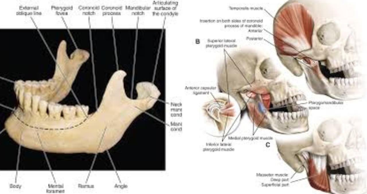
The neck of the mandibular condyle is the constricted portion just below the head, and this region is particularly important clinically as it is a common site of fracture. The neck is thinner than the head, making it a point of weakness when the jaw receives a blunt force impact. The lateral pole of the mandibular condyle is the attachment point for the lateral pterygoid muscle, a key muscle in the protrusion and lateral movement of the jaw. This muscle attaches to the pterygoid fovea, a small depression on the anterior aspect of the condylar neck. The medial pole, on the other hand, gives attachment to the sphenomandibular ligament. These attachments are crucial for the dynamic movement of the condyle, allowing it to slide forward and downward during mouth opening. The mandibular condyle does not exist in isolation; it is part of a larger functional unit that includes the teeth, muscles of mastication, and the temporomandibular joint capsule. Any alteration in the alignment of the teeth can affect the position of the mandibular condyle within the fossa, potentially leading to pain and dysfunction. Conversely, changes in the condyle, such as arthritis or fracture, can alter the bite, leading to a cascade of problems throughout the masticatory system.
Development and Growth of the Mandibular Condyle
The development of the mandibular condyle is a fascinating process that begins early in embryonic life. The mandible itself is the first bone of the face to ossify, and the condyle develops from a secondary cartilage that appears during the fetal period. This is in contrast to most other bones, which develop from a primary ossification center. The mandibular condyle is a significant growth center for the mandible, contributing to its length and height. This growth occurs through endochondral ossification, a process where cartilage is gradually replaced by bone. During childhood and adolescence, the mandibular condyle is highly active, allowing the jaw to grow in tandem with the cranium, accommodating the eruption of permanent teeth. This growth is not uniform; it is influenced by genetic factors, muscle function, and hormonal signals. The condylar cartilage acts as a growth plate, with cells proliferating and differentiating to form new bone, ultimately lengthening the ramus and body of the mandible.
The growth of the mandibular condyle is also responsive to functional demands, a phenomenon known as functional matrix theory. The soft tissues surrounding the bone, including the muscles and teeth, play a critical role in directing its growth. For instance, the forces of mastication stimulate the condylar cartilage, promoting new bone formation. If a person loses teeth on one side of their mouth, they may unconsciously chew more on the other side, leading to asymmetrical growth of the condyles. This adaptive capacity is what makes the mandibular condyle unique, but it also makes it vulnerable to developmental disturbances. Conditions like juvenile idiopathic arthritis can attack the condylar growth center, leading to a condition known as temporomandibular joint arthritis, which can result in a small, underdeveloped mandible, a condition called micrognathia. This can have profound aesthetic and functional consequences, affecting the patient’s breathing, swallowing, and self-esteem. Understanding the growth and development of the mandibular condyle is therefore essential for orthodontists and maxillofacial surgeons who treat children and adolescents with facial deformities.
The Role of the Mandibular Condyle in TMJ Function
The temporomandibular joint, or TMJ, is one of the most complex joints in the human body, and the mandibular condyle is its central figure. The TMJ is a ginglymo-arthrodial joint, meaning it allows for both hinge-like rotation and gliding movements. The rotational movement, which is the initial phase of opening the mouth, occurs between the mandibular condyle and the articular disc. This is the movement that allows us to open our mouths to about twenty-five millimeters. After this initial rotation, the condyle and disc translate forward together, sliding down the articular eminence of the temporal bone. This translation allows for wider mouth opening and is essential for powerful biting. The disc, a fibrous oval plate, acts as a shock absorber and stabilizer, ensuring that the condyle moves smoothly against the temporal bone. The integrity of the disc is crucial; if it becomes displaced, the condyle may click, pop, or lock, leading to significant discomfort and limitation of movement.
The mandibular condyle’s movement is dictated by a complex interplay of muscles, including the masseter, temporalis, medial pterygoid, and lateral pterygoid. The lateral pterygoid muscle is the primary protractor of the condyle, pulling it forward during mouth opening and in side-to-side grinding motions. The other muscles act as elevators, closing the jaw with incredible force. The muscles of mastication attach directly or indirectly to the mandible, and their coordinated contraction is orchestrated by the trigeminal nerve. Any disruption in this neuromuscular harmony, whether from stress, clenching, or trauma, can lead to a condition known as temporomandibular disorder, or TMD. TMDs are a cluster of disorders affecting the TMJ and the muscles of mastication, and they are a leading cause of chronic facial pain. The mandibular condyle is often at the center of these disorders, whether it is due to osteoarthritis, rheumatoid arthritis, disc displacement, or hypermobility. A thorough understanding of the condyle’s function is, therefore, the cornerstone of diagnosing and treating TMDs.
Imaging the Mandibular Condyle: A Window into Pathology
Given the critical role of the mandibular condyle in jaw function, accurate imaging is essential for diagnosis and treatment planning. Various imaging modalities are used to visualize the mandibular condyle, each with its own strengths and limitations. Panoramic radiography, or a panoramic X-ray, is often the first-line imaging technique in a dental or oral surgery clinic. It provides a broad overview of the mandible and the condyles, allowing the clinician to assess for gross abnormalities, such as fractures, tumors, or evidence of joint degeneration, like flattening or erosion of the condylar head. However, panoramic radiography provides a two-dimensional image and may not reveal subtle changes in the condyle or the joint space. It is a screening tool that, while valuable, often necessitates further, more detailed imaging for a definitive diagnosis.
For a more detailed view of the mandibular condyle, computed tomography, or CT, is the gold standard. CT scans provide cross-sectional images of the bone, offering an unparalleled view of the condyle’s internal architecture. They are particularly useful for assessing fractures, degenerative joint diseases, and bony ankylosis, a condition where the mandibular condyle fuses to the temporal bone. Cone beam CT, a specialized form of CT, is increasingly used in dental and maxillofacial practices because it provides high-resolution images of the condyle at a lower radiation dose than conventional CT. On the other hand, magnetic resonance imaging, or MRI, is the preferred imaging modality for evaluating the soft tissues of the TMJ, particularly the articular disc. MRI is the only imaging technique that can reliably visualize the position and morphology of the disc, and it can also detect early signs of inflammation or effusion in the joint. While CT excels at imaging bone, MRI excels at imaging the soft tissue structures that are often the source of pain in TMD patients. The combination of these imaging techniques provides a comprehensive picture of the mandibular condyle and its surrounding structures, allowing clinicians to tailor treatment to the specific pathology.
Fractures of the Mandibular Condyle: Causes, Diagnosis, and Management
Fractures of the mandibular condyle are among the most common injuries to the facial skeleton, accounting for a significant percentage of all mandibular fractures. These injuries are typically the result of blunt force trauma to the chin or the side of the face. The mechanism of injury is often a fall, a motor vehicle accident, or an assault. The condyle is a relatively slender part of the mandible, and when the jaw is forcefully impacted, the energy is transmitted through the mandibular body and ramus, concentrating at the condylar neck, which is the weakest point. Condylar fractures can be classified based on the level of the fracture—intracapsular, condylar neck, or subcondylar—and the degree of displacement. An intracapsular fracture involves the head of the condyle itself and can be particularly challenging to manage because it disrupts the articular surface. A condylar neck fracture occurs below the head but above the ramus, and a subcondylar fracture occurs at the base of the condylar neck.
The diagnosis of a mandibular condyle fracture is based on a thorough clinical examination and radiographic imaging. The patient will typically present with pain, swelling, and limited mouth opening. There may be an occlusal disturbance, meaning the patient’s bite feels off, and they may have an anterior open bite, where the front teeth no longer touch. In severe cases, the displaced condyle may cause bleeding from the ear canal if the fracture is high. Radiographic confirmation is essential, and panoramic radiography or CT scans are usually obtained. The management of condylar fractures has evolved significantly over the years. Historically, closed reduction, where the fracture is managed by wiring the jaws together, was the standard of care. Today, the trend is towards open reduction and internal fixation, where the fragments are surgically exposed and fixed with plates and screws. However, the treatment approach is highly individualized. In children, condylar fractures are often managed conservatively because of their remarkable ability to remodel and heal. In adults, the decision to operate depends on the degree of displacement, the patient’s age, and the presence of other injuries. For many nondisplaced or minimally displaced fractures, closed treatment with a period of rest and a soft diet is sufficient. The goal of treatment, regardless of the method, is to restore normal function and to prevent long-term complications, such as malocclusion, ankylosis, or chronic joint pain.
Degenerative Joint Diseases and the Mandibular Condyle
Degenerative joint disease, or osteoarthritis, is a common condition that can affect the mandibular condyle, leading to significant pain and functional impairment. Osteoarthritis of the TMJ is characterized by the progressive breakdown of the articular cartilage and the underlying bone of the condyle. It is often associated with aging, but it can also be caused by trauma, parafunctional habits like bruxism (teeth grinding), or a genetic predisposition. The pathophysiology involves a complex interplay of mechanical stress and inflammatory mediators. As the cartilage breaks down, the joint space narrows, and the underlying bone becomes exposed. The body attempts to repair this damage, leading to the formation of new bone, often in the form of osteophytes, or bone spurs, around the margins of the condyle. This leads to a characteristic radiographic appearance of the joint, with flattening of the condylar head, subchondral bone sclerosis (increased density), and osteophyte formation.
The clinical presentation of TMJ osteoarthritis is varied. Many patients may be asymptomatic, despite having significant radiographic changes. However, others may present with deep, aching pain in the joint, which is often worse with jaw function. They may experience crepitus, a grating or crackling sound in the joint during movement, which is caused by the roughened articular surfaces rubbing together. Morning stiffness is a common complaint, and the patient may have limited mouth opening. The management of TMJ osteoarthritis is primarily conservative, focusing on relieving symptoms and improving function. This includes patient education, a soft diet, the use of non-steroidal anti-inflammatory drugs to reduce pain and inflammation, and physical therapy. In some cases, occlusal splints, or bite guards, may be used to reduce the load on the joint by providing a stable occlusal platform. Corticosteroid injections into the joint can provide temporary relief, but they are not a long-term solution. For severe, refractory cases, surgical options, such as arthroscopy or joint replacement, may be considered, but these are reserved for the minority of patients. The management of TMJ osteoarthritis is a chronic, multi-modal process that requires a partnership between the patient and the healthcare provider.
Inflammatory Arthritis and the Mandibular Condyle
Inflammatory arthritides, such as rheumatoid arthritis, juvenile idiopathic arthritis, and psoriatic arthritis, also significantly impact the mandibular condyle. Unlike osteoarthritis, which is primarily a degenerative disease, inflammatory arthritis is caused by an autoimmune process that results in chronic inflammation of the synovial lining of the joint. This inflammation leads to the release of enzymes that break down cartilage and bone, eventually causing joint destruction. In the TMJ, the symptoms of inflammatory arthritis are often more severe than those of osteoarthritis. The pain is typically characterized as being more severe in the morning and after periods of rest, improving with activity. The joint is often warm and swollen, and there can be systemic signs of inflammation, such as fever and fatigue. In children with juvenile idiopathic arthritis, the mandibular condyle is commonly affected, which can have devastating consequences for facial growth.
The diagnosis of inflammatory TMJ arthritis is based on a combination of clinical findings, laboratory tests, and imaging. Blood tests for inflammatory markers, such as erythrocyte sedimentation rate and C-reactive protein, as well as specific antibodies like rheumatoid factor, are often elevated. Imaging studies, particularly CT and MRI, reveal the destructive changes in the condyle, including erosion, flattening, and, in severe cases, complete resorption of the condylar head. MRI can also reveal the presence of active synovitis and joint effusion. The treatment of inflammatory TMJ arthritis is focused on controlling the underlying systemic disease. This involves the use of disease-modifying antirheumatic drugs, or DMARDs, such as methotrexate, and biologic agents like TNF inhibitors. In the TMJ, intra-articular corticosteroid injections can be used to quickly reduce local inflammation. Physical therapy is also an essential component of care to maintain jaw mobility and prevent the development of fibrosis. In cases of severe growth disturbance in children, surgical intervention, such as a costochondral graft to reconstruct the condyle, may be needed. The management of inflammatory TMJ arthritis is complex and often requires a multidisciplinary approach involving rheumatologists, oral surgeons, and orthodontists.
Mandibular Condyle and Temporomandibular Disorders
The mandibular condyle is inextricably linked to temporomandibular disorders, a broad term that encompasses a variety of conditions affecting the TMJ, masticatory muscles, and associated structures. TMDs are a significant public health issue, affecting millions of people worldwide, and they are a leading cause of chronic orofacial pain. The etiology of TMDs is multifactorial, involving a complex interplay of genetic, psychological, and physiological factors. For many patients, stress and anxiety play a significant role, leading to muscle hyperactivity and clenching, which overloads the joint. Parafunctional habits, such as bruxism or nail-biting, are also major contributing factors. Trauma, either from a single event or from repetitive microtrauma, can precipitate or exacerbate TMD symptoms. The mandibular condyle is often the primary site of pathology in TMDs, with conditions like disc displacement with reduction (clicking or popping), disc displacement without reduction (locked jaw), and osteoarthritis being the most common.
The signs and symptoms of TMDs are varied, but they typically include pain in the jaw joint, face, or ears, which is often worse with chewing or talking. Patients may experience a limited range of motion in the jaw, or they may have a sensation that the joint is catching or locking. Joint sounds, such as clicking, popping, or crepitus, are also common. Many patients also experience headache, neck pain, and tinnitus. The diagnosis of TMDs is primarily clinical, based on a thorough history and physical examination. Imaging may be used to confirm the diagnosis or to rule out other causes, but it is not always necessary. The management of TMDs is conservative and multidisciplinary. For most patients, the focus is on self-management and behavioral interventions. This includes patient education, jaw rest, a soft diet, the application of heat or cold, and relaxation techniques. Occlusal splints, or bite guards, are commonly used to reduce the load on the joint and to help break the cycle of parafunctional activity. Physical therapy, including exercises to strengthen the jaw muscles and improve coordination, is also beneficial. In more severe cases, pharmacological management, including the use of NSAIDs, muscle relaxants, or low-dose antidepressants for pain management, may be indicated. For a subset of patients who do not respond to conservative therapy, more invasive procedures, such as arthrocentesis, arthroscopy, or open joint surgery, may be considered. However, surgery is generally viewed as a last resort due to its risks and variable outcomes.
Surgical Interventions and the Mandibular Condyle
Surgery on the mandibular condyle is a highly specialized area of oral and maxillofacial surgery, and it is reserved for conditions that do not respond to conservative therapy. The indications for surgery include severe, intractable joint pain, a locked jaw that cannot be reduced (closed lock), significant dysfunction that limits quality of life, and advanced degenerative or inflammatory joint disease. Surgical procedures on the condyle can be broadly classified into three categories: arthroscopic surgery, open joint surgery, and joint replacement. Arthroscopy is a minimally invasive procedure that involves inserting a small camera and instruments into the joint through small incisions. It is used to diagnose joint pathology, remove adhesions (scar tissue), reposition the disc, or remove loose bodies. Arthroscopy is typically an outpatient procedure and is associated with fewer complications and a faster recovery compared to open surgery. It is a valuable tool for managing patients with disc displacement and early arthritis.
Open joint surgery, or arthrotomy, involves directly exposing the joint through a larger incision. This is a more invasive procedure and is used for more complex conditions. During an arthrotomy, the surgeon can perform a variety of procedures, including disc repositioning, discectomy (removal of the disc), and condylar shave (recontouring the condyle). Another common open joint procedure is the eminectomy, where the articular eminence is lowered to prevent the condyle from being locked in a forward position. In severe cases of joint destruction, such as advanced arthritis or ankylosis, total joint replacement may be necessary. This involves replacing the entire mandibular condyle and the glenoid fossa with custom-made, biocompatible implants. Total joint replacement is a major operation, but it can be a life-changing procedure for patients who have exhausted all other treatment options, providing significant pain relief and restoring jaw function. The choice of surgical intervention is highly individualized and depends on the specific diagnosis, the patient’s age and overall health, and their expectations. The decision to proceed with surgery should only be made after a careful discussion between the patient and the surgeon about the risks, benefits, and alternatives.
Trauma and the Mandibular Condyle: Managing Acute Injuries
Acute trauma to the mandibular condyle, as discussed, is a common clinical scenario. Beyond fractures, trauma can also result in dislocation of the condyle. A dislocation occurs when the mandibular condyle moves too far forward and becomes stuck anterior to the articular eminence. This is a painful condition that prevents the patient from closing their mouth. The dislocation can be caused by a traumatic blow, but it is often triggered by a wide yawn, a dental procedure, or an episode of vomiting. When the patient presents with an open bite that they cannot close, the diagnosis is often straightforward. The acute management of a dislocation involves manually reducing the condyle back into its fossa. This is a procedure that requires a significant amount of strength and skill, and it is usually performed in an emergency setting. The physician or dentist will position their thumbs on the lower molars, apply downward pressure, and push the mandible backward until the condyle slips back into place. This can be a painful procedure, and patients are often given sedation or local anesthesia to help them tolerate it. After a dislocation, the patient is typically placed on a soft diet and advised to avoid wide mouth opening for several weeks to allow the tissues to heal and to prevent recurrence.
The management of acute trauma to the mandibular condyle also requires a thorough assessment for other injuries. Because condylar fractures are often the result of high-energy impacts, there is a risk of concurrent injuries to the cervical spine, the brain, or other facial bones. The healthcare provider must perform a complete trauma evaluation, including a neurological examination and imaging of the cervical spine. In the case of condylar fractures, the immediate priority is to stabilize the patient and ensure their airway is secure. Once the patient is stabilized, a definitive treatment plan for the fracture can be formulated. Early intervention in acute condylar injuries is crucial to prevent long-term complications, such as malocclusion, chronic pain, and ankylosis. The acute phase of injury is also a critical time for patient education. The patient should be counseled on what to expect during the recovery process and how to manage their symptoms. This includes advice on diet, pain management, and follow-up care. The psychological impact of facial trauma should not be underestimated, as many patients experience anxiety and distress. A compassionate and supportive approach from the healthcare team is essential for the patient’s overall well-being.
The Mandibular Condyle and Orthognathic Surgery
The mandibular condyle plays a pivotal role in orthognathic surgery, which is surgery to correct deformities of the jaws and face. These deformities can be congenital, such as in patients with cleft lip and palate, or acquired, as a result of trauma, disease, or developmental disturbances. The goal of orthognathic surgery is to reposition the jaws to improve function, aesthetics, and stability. When planning a surgical procedure on the mandible, such as a sagittal split ramus osteotomy, the surgeon must pay careful attention to the position of the mandibular condyle. The sagittal split osteotomy involves cutting the ramus of the mandible and moving the body of the mandible forward or backward to correct the bite. The condyle is left attached to the proximal segment, which is then repositioned and secured to the distal segment with plates and screws. If the condyle is not properly positioned in the fossa during the procedure, the patient can experience postoperative complications, including TMJ pain, limited mouth opening, and condylar resorption.
The position of the mandibular condyle after orthognathic surgery is a topic of ongoing research and debate. Surgeons have developed various techniques to ensure the condyle is seated in its optimal position at the end of the procedure. Some surgeons use manual manipulation, while others use specialized instruments, such as a condylar positioner, to guide the condyle into place. Advances in imaging technology, including intraoperative CT, have also been used to verify the condyle’s position before the patient leaves the operating room. The importance of accurate condylar positioning cannot be overstated; it is a key determinant of the success of the surgery and the stability of the outcome. If the condyle is displaced or rotated, it can lead to an unstable occlusion and the potential for relapse, where the jaw moves back to its original position. Long-term follow-up of orthognathic surgery patients has shown that even minor changes in condylar position can have significant consequences over time, leading to degenerative joint changes and the need for additional surgery. Therefore, the mandibular condyle is not just a passive structure in orthognathic surgery; it is an active participant that must be carefully managed to achieve a successful result.
The Mandibular Condyle and Obstructive Sleep Apnea
Obstructive sleep apnea is a serious condition where the airway collapses during sleep, leading to repeated pauses in breathing. While the mandibular condyle is not the primary cause of OSA, it is a critical component in one of its most common treatments: mandibular advancement devices. These oral appliances work by holding the mandible in a forward position, which in turn pulls the tongue and the soft palate forward, opening the airway. The mandibular condyle is crucial to the success and safety of these devices. When the mandible is advanced, the condyle is translated forward and downward along the articular eminence. The device must hold the jaw in this position comfortably for the duration of the sleep period, without causing strain on the condyle or the TMJ. In patients with TMD or joint pain, a mandibular advancement device can exacerbate their symptoms, making it essential for the treating physician to assess the patient’s TMJ health before prescribing such a device.
Furthermore, the long-term use of a mandibular advancement device can have effects on the mandibular condyle and the TMJ. Over time, the sustained forward positioning of the condyle can lead to remodeling of the bony structures, and in some cases, it can cause progressive changes in the occlusal relationship. Patients who use these devices for many years may notice that their bite changes, with their front teeth no longer touching. This is because the condyle, over time, adapts to the new position, and the posterior teeth may supraerupt. This is a well-documented phenomenon, and it underscores the importance of regular dental monitoring for patients using mandibular advancement devices. The management of OSA with oral appliances is a delicate balance between effectively managing the airway and preserving the long-term health of the TMJ and the mandibular condyle. The ideal device provides effective airway management while minimizing the load on the condyle. In recent years, there has been a trend towards more sophisticated devices that are titratable, allowing the patient to adjust the degree of mandibular advancement to achieve the optimal therapeutic effect with the least side effects.
Rehabilitation and Physical Therapy for the Mandibular Condyle
When the mandibular condyle is affected by injury or disease, rehabilitation and physical therapy are essential components of the recovery process. The primary goal of physical therapy is to restore normal function to the TMJ and to reduce pain. This is achieved through a combination of manual therapy, therapeutic exercises, and patient education. Manual therapy involves hands-on techniques performed by a trained therapist to improve joint mobility, reduce muscle tension, and decrease pain. This may include joint mobilization, where the therapist gently manipulates the condyle to improve its range of motion, or soft tissue massage to relieve tension in the muscles of mastication. Physical therapy also focuses on correcting muscle imbalances, as muscle dysfunction is often a contributing factor to TMDs.
Therapeutic exercises are a cornerstone of TMJ rehabilitation. These exercises are designed to strengthen the muscles that open the mouth, improve coordination, and retrain the jaw to move in a more symmetric and functional pattern. For example, a common exercise involves placing the tongue on the roof of the mouth and slowly opening and closing the mouth. This helps to limit the range of motion and prevents the jaw from locking. Another exercise involves using a finger to resist the movement of the jaw, strengthening the muscles. It is crucial that patients are taught these exercises correctly to prevent further injury. Patient education is another critical aspect of rehabilitation. Patients need to understand the nature of their condition and learn how to manage it independently. This includes learning to identify and avoid triggers, such as hard or chewy foods, and to adopt relaxation techniques to reduce stress and muscle tension. A successful rehabilitation program empowers the patient to take control of their condition, leading to a long-term improvement in their quality of life. The mandibular condyle, like any other joint, thrives on controlled, low-load movement, and rehabilitation is the key to achieving this.
The Future of Mandibular Condyle Research
The field of mandibular condyle research is rapidly evolving, with exciting advances on the horizon. One of the most promising areas of research is in tissue engineering and regenerative medicine. Scientists are exploring ways to grow new cartilage and bone to repair or replace damaged condyles. This is particularly relevant for patients with severe arthritis or those who have had their condyles removed due to a tumor. Current treatment options for these patients are limited, and they often involve complex reconstructive surgeries. Tissue engineering offers the hope of a more biological and less invasive solution. Researchers have been successful in growing condylar cartilage in the laboratory using stem cells and biomaterials, and they are now working towards translating this research to human patients. The potential of this approach is enormous, as it could revolutionize the treatment of TMJ disorders and significantly improve the quality of life for millions of people.
Another area of active research is the biomechanics of the mandibular condyle. Using advanced computer models and simulation techniques, researchers are gaining a better understanding of the complex forces that act on the condyle during function. This knowledge is being used to design better diagnostic tools and to develop more targeted treatment strategies. For example, by understanding the stress distribution within the condyle, researchers can predict the risk of fracture or the progression of arthritis. This can help clinicians identify patients who are at high risk of developing TMDs and to intervene earlier. There is also a growing interest in the role of genetics in condylar development and disease. By identifying the genes that are involved in condylar growth and degeneration, scientists hope to develop new therapies that can prevent or halt the disease process. The mandibular condyle, once a relatively obscure anatomical structure, is now a focal point of biomedical research. The future holds the promise of new diagnostic tools, more effective treatments, and ultimately, a better understanding of this remarkable and vital joint. The integration of artificial intelligence and machine learning into radiographic analysis is also poised to improve the accuracy of diagnosing condylar pathologies, allowing for more precise and personalized treatment plans. As our understanding of the mandibular condyle deepens, so too will our ability to alleviate the pain and suffering associated with its dysfunction.